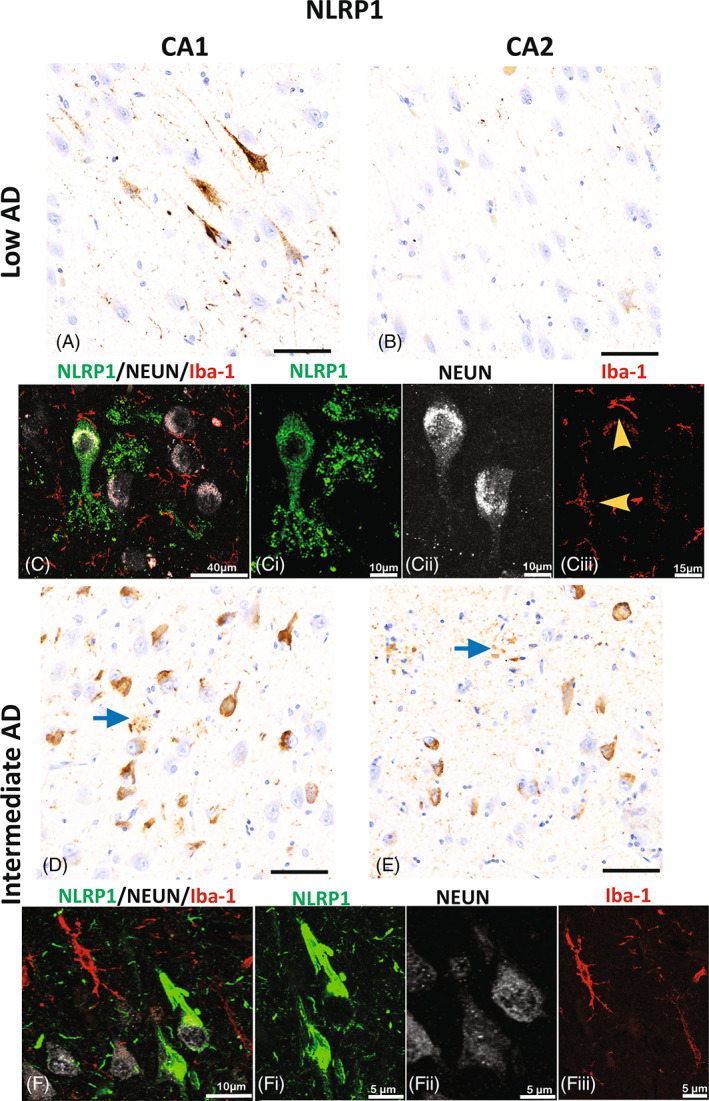
FIGURE 4.

NLRP1 expression in hippocampal neurons. Immunoreactivity of NLRP1 in the CA1 (A and D) and CA2 (B and E) hippocampal regions in low AD (A–C) and in intermediate AD (D–F) cases. Expression of NLRP1 is present in neurons and apical dendrites in the CA1 region of low AD (A) more than in the CA2 (B) hippocampal region. In CA1, the cytoplasmic neuronal expression of NLRP1 (green, C and Ci) is confirmed by the confocal photomicrographs that show the expression in neurons (white; Cii), but not in microglia (yellow arrow heads; red; Ciii). In the intermediate AD, NLRP1 immunoreactivity is seen in numerous neurons and in parenchyma in the form of clusters (blue arrows) of the CA1 (D) and CA2 (E). The photomicrographs capture the dense neuronal cytoplasmic expression seen in CA1 of the intermediate AD cases (F and Fi) which is not present in the microglia (red; Fiii). Alzheimer's disease (AD); NOD‐like receptor proteins (NLRP); Cornu Ammonis Field 2 (CA2); Cornu Ammonis Field 1 (CA1); images A, B, C, and E; scale bar = 60 μm.
