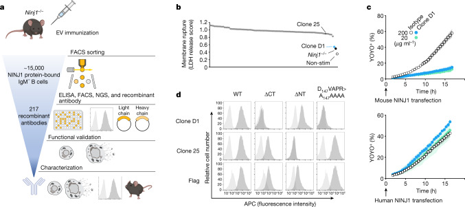
Fig. 1. Identification of NINJ1-blocking antibody clone D1.
a, Scheme of recombinant antibody screening. EV, extracellular vesicle; FACS, fluorescence-activated cell sorting. b, LDH released from Pam3CSK4-primed wild-type BMDMs after nigericin stimulation for 16 h in the presence of 1 μg ml−1 of indicated antibody (dots represent different antibodies tested). Ninj1−/−, Ninj1−/− BMDMs; non-stim, non-stimulated wild-type BMDMs. The LDH score is the LDH release normalized against the no-antibody control. c, The percentage of YOYO-1+ NINJ1-expressing HEK293T cells when cultured with clone D1 or an isotype control antibody. Data are mean (circles) ± s.d. (shaded area) of three independent replicates. d, Flow cytometry histograms of propidium iodide-negative HEK293T cells surface-stained with anti-NINJ1 or anti-Flag antibodies. Cells are mock-transfected (light grey) or transfected with indicated NINJ1 constructs (dark grey). WT, wild type; ΔCT, NINJ1(Δ142–152); ΔNT, NINJ1(Δ2–73). In c,d, results are representative of three independent experiments.

