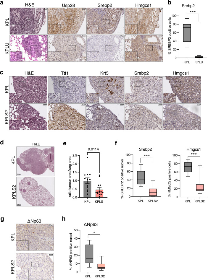
Fig. 6. Deletion of Srebf2 attenuates tumour formation in a mouse model of lung squamous cell carcinoma.
a Tissue sections from KPL and KPLU lung tumours were stained for Usp28, Srebp2 and Hmgcs1 by immunohistochemistry. Haematoxylin and eosin (H&E) staining is also shown. b Boxplot showing quantification of Srebp2 staining in KPL and KPLU tumours. Percent positive cells are shown. (KPL: n = 9; KPLU: n = 7; ***p < 0.001, Mann–Whitney test). c Tissue sections from KPL and KPLS2 tumours were stained for the adenocarcinoma marker thyroid transcription factor 1/NK2-homeobox 1 (Ttf-1/Nkx2-1), the squamous marker keratin 5 (Krt5), Srebp2 and Hmgcs1 by immunohistochemistry. H&E staining is also shown. d H&E staining of representative lung tissue sections from KPL and KPLS2 mice. e Ratio of tumour area relative to total lung area in KPL and KPLS2 mice. Data are displayed as mean ± SD (KPL: n = 11; KPLS2: n = 13; Mann-Whitney test). f Boxplot showing quantification of Srebp2 and Hmgcs1 staining in KPL and KPLS2 tumours. Percent positive nuclei or positive cells are shown. (KPL: n = 7; KPLS2: n = 9; ***p < 0.001, Mann-Whitney test). g Tissue sections from KPL and KPLS2 tumours were stained for ΔNp63. h Boxplot showing quantification of ΔNp63 staining in KPL and KPLS2 tumours. Percent positive nuclei are shown. (KPL: n = 6; KPLS2: n = 6; ***p < 0.05, Mann-Whitney test).

