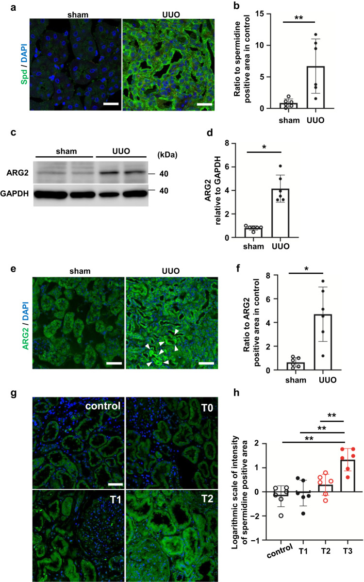
Fig. 2. UUO increases spermidine and ARG2 protein levels in the mouse kidney.
a Confocal immunofluorescence microscopic images of Spd in control and UUO kidneys. Green, anti-Spd antibody; blue, DAPI. Scale bars, 50 µm. b Quantification of the Spd-positive area in control and UUO kidneys (n = 6 in each group). Data are indicated as means ± SD. c Western blot analysis of ARG2 protein levels in the whole kidney of sham and UUO mice. d Relative levels of ARG2 protein normalized to GAPDH in sham and UUO kidneys are shown (n = 6 in each group). Data are indicated as means ± SD. e Immunohistochemistry of ARG2 in control and UUO kidneys. Green, anti-ARG2 antibody; blue, DAPI. Scale bars, 50 µm. White arrowheads indicate ARG2-positive tubules. f Quantification of the ARG2-positive area in sham and UUO kidneys (n = 6 in each group). Data are indicated as means ± SD. *P < 0.05, **P < 0.01. g Immunostaining of Spd in the human kidney. Control, donor kidneys; T0–T2, interstitial fibrosis/tubular atrophy scores from the Oxford classification of IgA nephropathy. The percentages of lesions in the cortical area were as follows: T0 = 0%–25%, T1 = 26%–50%, and T2 = > 50%. Scale bars, 50 µm. h Quantification of the Spd-positive area in human kidneys (n = 6 in each group). Data are indicated as means ± SD. **P < 0.01. Spd spermidine, UUO unilateral ureteral obstruction, ARG2 arginase 2, DAPI 4′,6-diamidino-2-phenylindole, GAPDH glyceraldehyde-3-phosphate dehydrogenase.

