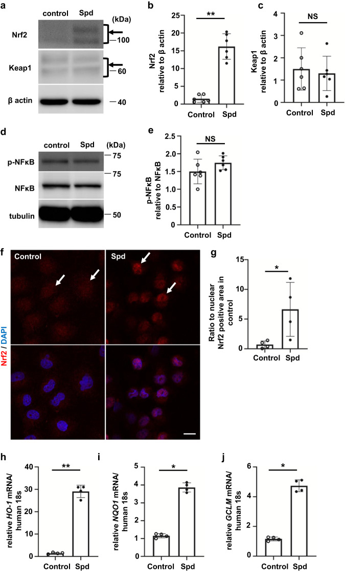
Fig. 4. Spd activates the transcription factor Nrf2 in renal tubular epithelial cells.
a Western blot analysis of Nrf2 and Keap1 protein levels in HK-2 cells incubated with Spd. The arrow shows two Nrf2 bands. b Relative levels of Nrf2 protein normalized to β-actin are shown (n = 6 in each group). Data are indicated as means ± SD. c Relative levels of Keap1 protein normalized to β-actin are shown (n = 6 in each group). Data are indicated as means ± SD. d Western blot analysis of phospho- and total NF-κB in HK-2 cells incubated with Spd. e Relative levels of phospho-NF-κB protein normalized to total NF-κB are shown (n = 6 in each group). Data are indicated as means ± SD. f Immunofluorescence images of Nrf2 in HK-2 cells incubated with Spd. Red, anti-Nrf2 antibody; blue, DAPI. Scale bars, 20 µm. g Quantification of the nuclear Nrf2-positive area corrected for nuclei. Data are indicated as means ± SD. h HO-1, i NQO1, and j GCLM mRNA expression determined by real-time PCR in HK-2 cells incubated with Spd (n = 4 in each group). Data are indicated as means ± SD. *P < 0.05, **P < 0.01. Spd spermidine, Nrf2 nuclear factor erythroid 2-related factor 2, Keap1 kelch-like ECH-associated protein 1, NF-κB nuclear factor-κB, DAPI 4′,6-diamidino-2-phenylindole.

