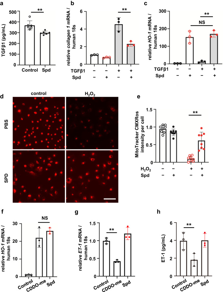
Fig. 6. Spd suppresses fibrotic signaling, but does not inhibit the endothelin pathway like bardoxolone methyl.
a Secretion of TGFβ1 from HK-2 cells treated with Spd measured by ELISA (n = 6 in each group). Data are indicated as means ± SD. b Collagen 1 and c HO-1 mRNA expression determined by real-time PCR in HK-2 cells incubated with TGFβ1 in the presence of Spd (n = 3 in each group). Data are indicated as means ± SD. d MitoTracker Red CMXRos staining in HK-2 cells when exposed to H2O2 and Spd. Scale bar, 100 µm. e Quantification of Mitotracker Red CMXRos intensity measured by a multimode plate reader (n = 8 in each group). Data are indicated as means ± SD. f HO-1 and g ET-1 mRNA expression determined by real-time PCR in HK-2 cells incubated with Spd and CDDO-me (n = 3 in each group). Data are indicated as means ± SD. h Secretion of ET-1 from HK-2 cells treated with Spd and CDDO-me measured by ELISA (n = 3 in each group). Data are indicated as means ± SD. **P < 0.01. NS not significant, Spd spermidine, TGFβ1 transforming growth factor β1, CDDO-me bardoxolone methyl.

