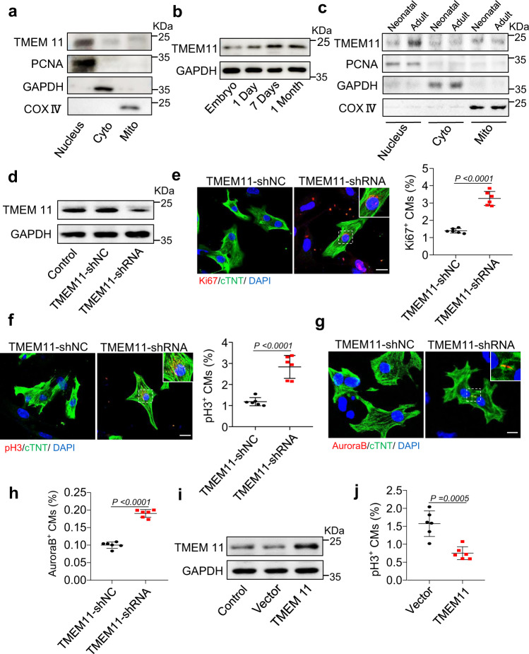
Fig. 1. TMEM11 participates in the regulation of proliferation in cardiomyocytes.
a Representative western blots showing the expression of TMEM11 in the nucleus, cytoplasm and mitochondria of cardiomyocytes (n = 3 independent experiments). b Representative western blots showing the expression of TMEM11 in hearts at different ages of mice (n = 4 mice). c Representative western blots showing the expression of TMEM11 in the nucleus, cytoplasm and mitochondria of cardiomyocytes (n = 3 mice). d–h Neonatal mice cardiomyocytes were infected with adenovirus harboring TMEM11-shRNA or its control (TMEM11-shNC) for 48 h. d The expression levels of TMEM11 mRNA assessed by immunoblot (n = 3 independent experiments). e Representative confocal images of Ki67-positive cardiomyocytes (Bar = 20 μm) and quantification of Ki67-positive (right) cells (n = 6 independent experiments). f Representative confocal images of pH3-positive cardiomyocytes and quantification of pH3-positive cells in TMEM11-shNC and TMEM11-shRNA infected cardiomyocytes (Bar = 20 μm) (n = 6 independent experiments). Representative confocal images of Aurora-B-positive cardiomyocytes (g) and quantification (h) of Aurora-B-positive cells in TMEM11-shNC and TMEM11-shRNA infected cardiomyocytes (Bar = 20 μm) (n = 6 independent experiments). i, j Neonatal mice cardiomyocytes were infected with adenovirus harboring negative control (Vector) or mouse TMEM11 gene (TMEM11) for 48 h. i Representative western blots showing the expression of TMEM11 (n = 4 independent experiment). j PH3-positive cardiomyocytes were measured in Vector and TMEM11 treated cardiomyocytes (n = 6 independent experiment). Data are presented as the mean ± s.d. Two-sided Student’s t test (e, f, h and j) was performed.

