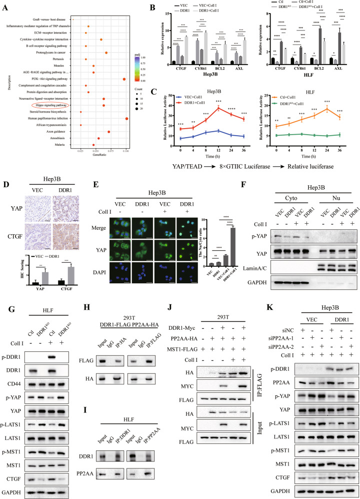
Fig. 6. DDR1 impedes Hippo signaling through inactivating YAP.
A KEGG pathway enrichment analysis indicates that the Hippo signaling pathway was significantly affected by HLF-shDDR1 cells treated with collagen I. B mRNA expression levels of Hippo pathway downstream-associated genes in DDR1 overexpression or knock-out HCC cell lines. C Luciferase reporter assays in DDR1 overexpression or knock-out HCC cells transfected with YAP/TAZ-responsive synthetic promoter-reporter plasmids and collagen I treatment. D Representative images of IHC staining of YAP and CTGF from BALB/c nude mice xenograft (scale bar = 100 μm). E Representative images of immunofluorescence assays display nucleus/cytoplasm localization of YAP in DDR1 overexpressed Hep3B cells with collagen I (scale bar = 30 μm). F Nucleus/cytoplasm fractionation and western blot analysis of DDR1 overexpressed cells to show YAP nuclear translocation. GAPDH and Lamin A/C were used as cytoplasmic and nuclear markers, respectively. G Effects of protein expression or phosphorylation of Hippo signaling components in DDR1 knock-out HLF cells with collagen I. H Co-IP and western blot assays for exogenously proteins DDR1-FLAG and PP2AA-HA were performed in HEK-293T cells. Cross-validation by immunoprecipitation with anti-HA, immunoblotting with anti- FLAG, immunoprecipitation with anti-FLAG, and immunoblotting with anti-HA. I Co-IP and western blot assays for endogenously proteins DDR1 and PP2AA were performed in HLF cells. Cross-validation by immunoprecipitation with anti-CD44, immunoblotting with anti-DDR1, immunoprecipitation with anti-DDR1, and immunoblotting with anti- PP2AA. J DDR1-Myc, PP2AA-HA, and MST1- FLAG were simultaneously transfected in HEK-293T cells with or without collagen I treatment, and evaluate the collagen I contributed to the indicated protein interaction and stability by western blot. K DDR1 overexpressed cells transfected with siPP2AA or control siRNA in the presence of collagen I treatment. Western blot analyzed the indicated protein from cell lysate. Two-tailed unpaired Student’s t-test was performed. Each bar represents the mean ± SD. *p < 0.05, **p < 0.01 and ***p < 0.001.

