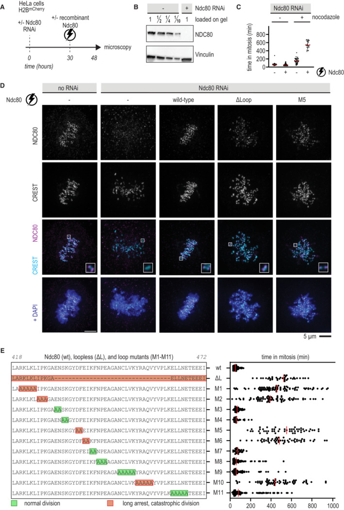
Figure 4. Mutation of critical residues in the loop impairs chromosome congression.

- Schematic of an electroporation experiment.
- Immunoblot of NDC80 levels following depletion of the Ndc80 complex by RNAi.
- Quantification of the time that cells spent in mitosis following various treatments. Each dot represents a cell and the red lines indicate median values. Nocodazole was added 17 h after electroporation and 1 h before microscopy. A minimum of 30 cells were analyzed for each condition.
- Immunofluorescence microscopy of mitotic cells stained for DNA (DAPI), kinetochores (CREST), and Ndc80 complexes. The Ndc80 antibody (9G3) detects endogenous Ndc80 (column 1) and electroporated recombinant Ndc80 (columns 3, 4, 5). Representative cells with a metaphase plate or uncongressed chromosomes are shown. Scale bar: 5 μm.
- Overview of mutations in the Ndc80 loop region and the effects on the time spent in mitosis following the experimental setup outlined in panel (A). Colors indicate whether cells divided normally, sometimes with delayed chromosome congression (green), or showed long arrests followed by a catastrophic division (orange). Each dot represents a cell and the red lines indicate median values. Mutation NDC80G434A‐Y435A did not support the formation of stable and soluble Ndc80 complexes. A minimum of 30 cells were analyzed for each condition.
Source data are available online for this figure.
