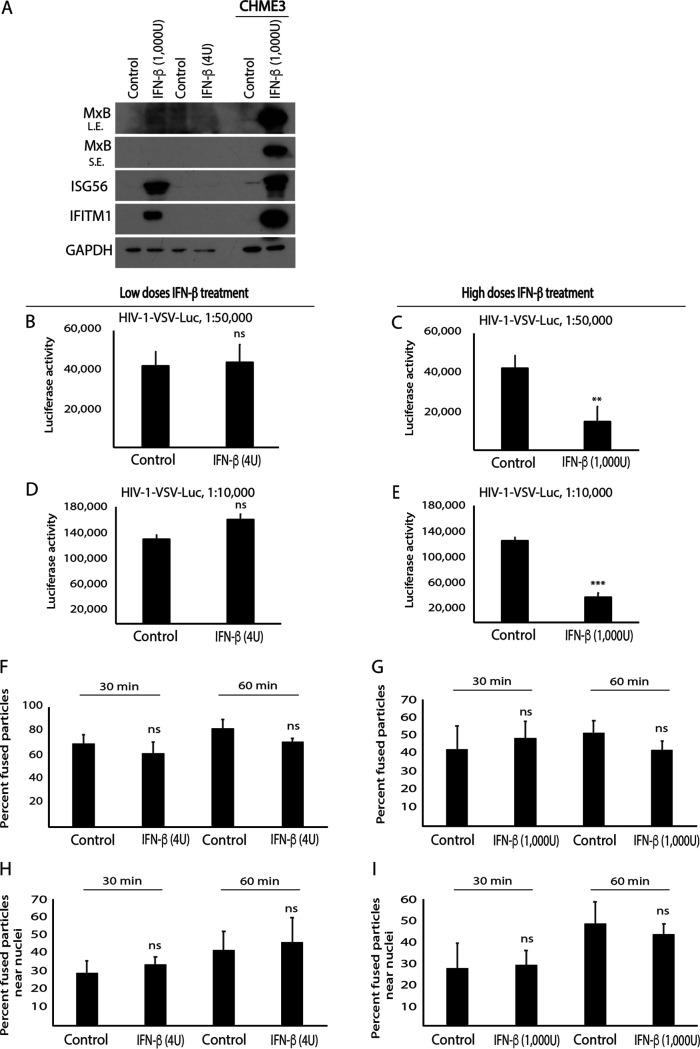
FIG 3.
IFN-β does not affect HIV-1 translocation to the nucleus in HEK293A cells. (A) WB analysis showing ISG (MxB, ISG56, and IFITM1) levels in HEK293A cells treated with various concentrations of IFN-β (low, 4 U; high, 1,000 U). Control or IFN-β treated CHME3 cells were included as the positive control for ISG induction. L.E., long exposure; S.E., short exposure. (B to I) HEK293A cells treated with low (B and D) or high (C and E) IFN-β concentrations were infected either with HIV-1 carrying a luciferase reporter and pseudotyped with VSV envelope (HIV-1-VSV-luc) at two different dilutions as indicated followed by measurements of luciferase activity (B–E) or with double-labeled HIV-1 (S15-tomato/GFP-Vpr) carrying a VSV envelope (HIV-1-DL-VSV) followed by measurements of the number of fused particles (S15-tomato−/GFP-Vpr+) in cells fixed at the indicated time postinfection (F–I). The percentage of double-labeled HIV-1-DL-VSV fused into the cytosol (F and G) or their accumulation within 2 μm of the nucleus (H and I) is shown. Data in panels F to I represent the mean of ≥730 virus particles analyzed in ≥55 fields of view (≥60 cells) for each condition. Data are shown as mean ± SD. Statistical significance was calculated using a t test (B to I). **, P ≤ 0.01; ***, P ≤ 0.001; ns, not significant. Results in panels A to E are representative of 3 and in panels F to I of 2 independent biological repeats.

