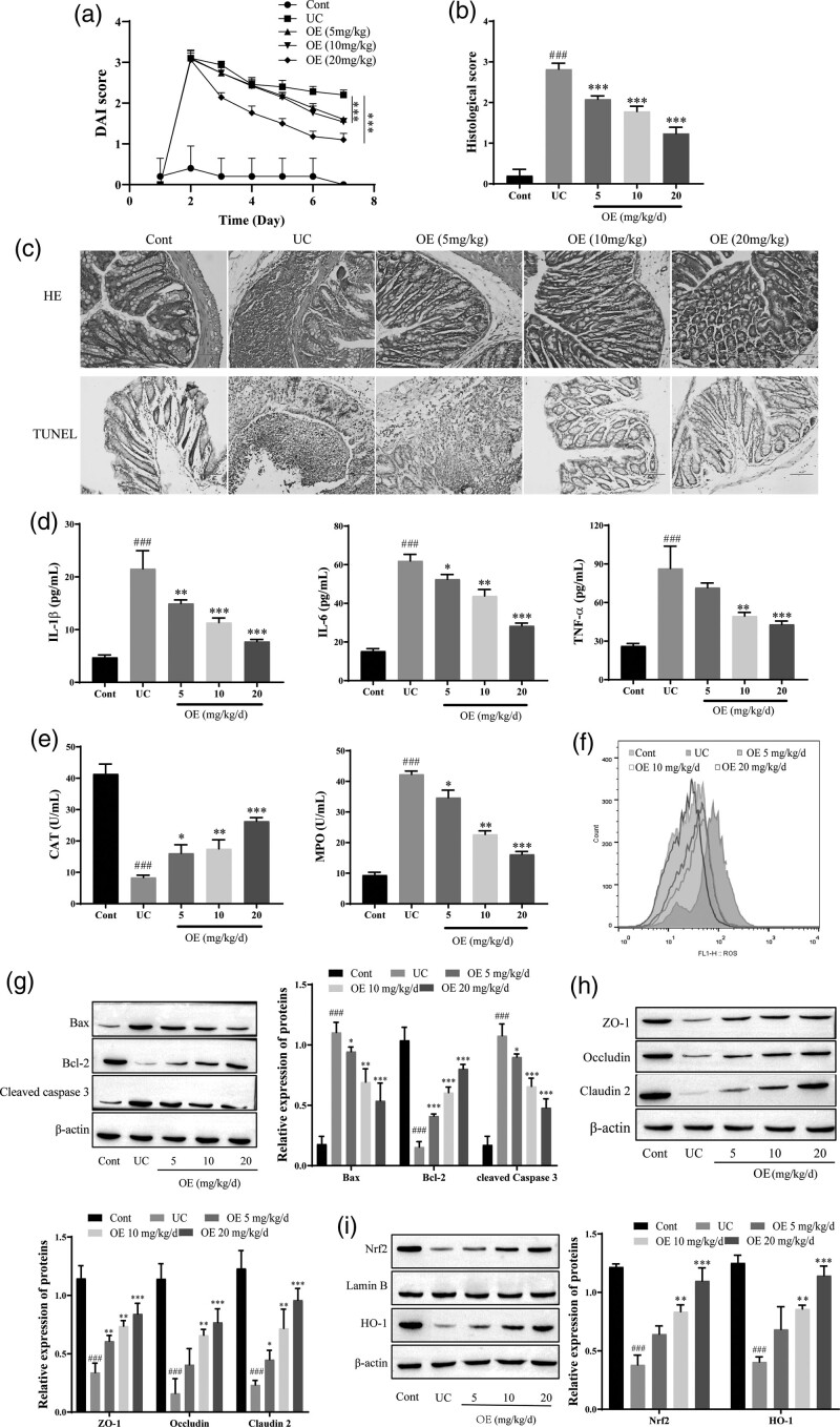
Fig. 1.
Effect of OE on TNBS-induced increases in tissue levels of inflammatory factors, intestinal epithelial cell apoptosis, and oxidative stress levels in UC rats. (a) Disease activity index (DAI) scores. (b) HE staining and histological score for the degree of damage in the rat colon (scale bar = 100 µm). (c) TUNEL for histomorphological changes in rats with UC (scale bar = 100 µm). (d) ELISA for IL-6, IL-1β, and TNF-α. (e) The contents of MPO and CAT were determined by kits. (f) Flow cytometry analysis of ROS levels. (g) Western blots showing the levels of Bax, Bcl2, and cleaved caspase 3. (h) Western blots showing the levels of the tight junction proteins ZO-1, Occludin, and Claudin-2. (i) Western blots showing the levels of Nrf2 and HO-1. Compared with the NC group, ###P ≤ 0.001; Compared with the UC group, *P ≤ 0.05, **P ≤ 0.01, ***P ≤ 0.001.

