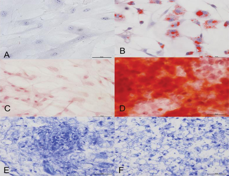
Fig. 2.

(A-F) Differentiation of mesenchymal cells. (B) The differentiation into the adipocyte lineage was demonstrated by staining with Oil Red O; (D) Alizarin Red S staining shows mineralization of the extracellular matrix in the osteogenic differentiation; and (F) toluidine blue shows the deposition of proteoglycans and lacunae in the chondrogenic differentiation. (A, C, E) Untreated control cultures without adipogenic, osteogenic or chondrogenic differentiation stimuli are shown.
