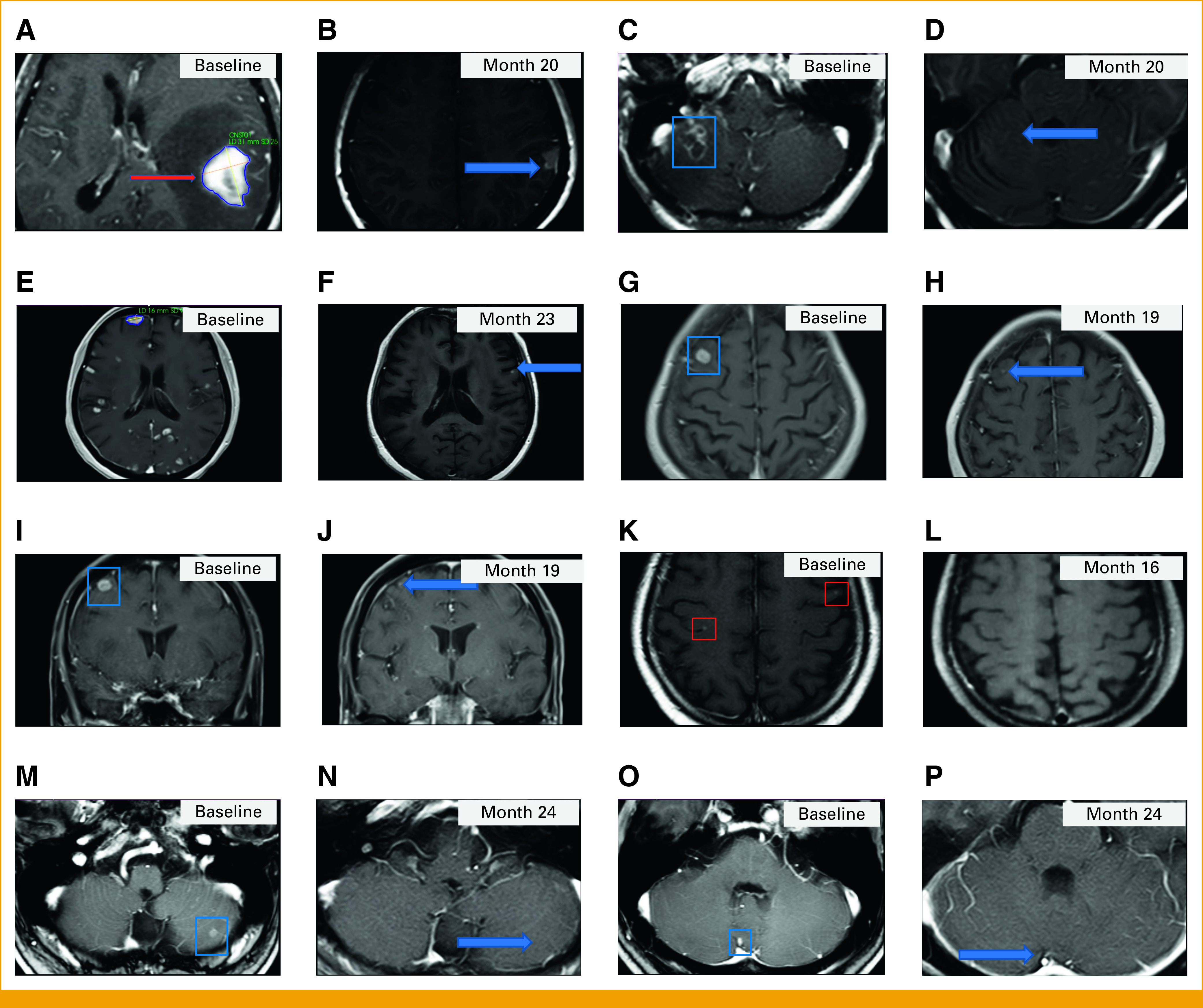
FIG 2.

Postcontrast T1-weighted MRI brain images. Patient 1: A 31 mm × 25 mm enhancing left parietal lesion (A) at baseline (red arrow) that was absent at month 2 and still absent (B) at month 20 (the blue arrow shows a hyperintense cavity) and an enhancing right cerebellar lesion (C) at baseline that was absent from month 8 and still absent (D) at month 20 (the blue arrow shows the expected location of the lesion). Patient 2: Multiple brain metastases (E) at baseline and a decrease in size and number of the enhancing brain metastases (F) at month 23, including resolution of the enhancing right frontal lesion. The blue arrow in (F) points to a tiny enhancing lesion. Patient 3: (G) Axial and (I) coronal baseline images demonstrating a 13 mm × 10 mm enhancing right frontal lesion and demonstrating a decrease in size of the right frontal lesion compared with baseline (H and J) at month 19. Blue arrows in (H) and (J) point to the smaller on study enhancing right frontal lesion. Patient 7: A small bilateral frontal enhancing lesions (K) at baseline demonstrating resolution (L) at month 16. Patient 8: Enhancing left cerebellar lesions (M and O) at baseline that were absent from month 2 and 10, respectively, with no visible lesions in the same locations at (N and P) month 24 (blue arrows show the expected locations of the lesions). The same T1-weighted imaging sequence was used to acquire all images. MRI, magnetic resonance imaging.
