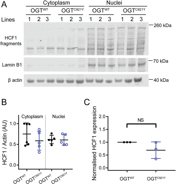
Fig. 4.

HCF1 processing by undifferentiated OGTWT and OGTC921Y in mESCs. (A) Immunoblot of HCF1 proteolytic fragments in the cytoplasm and nucleus of mESCs harbouring either OGTWT or the OGTC921Y variant. Lamin B1 was used as a marker for the nuclear fraction. This experiment was performed using three cell clones per genotype and repeated over two passages. (B) Quantification of HCF1 signal. HCF1 signals were normalised to those of β-actin. n=6 biological replicates. One-way ANOVA with Tukey’s comparison test; cytoplasmic fraction, P=0.99; nuclear fraction, P=0.54. (C) RT-PCR analysis of Hcf1 mRNA expression levels normalised to those of Gapdh, 18S (Rn18s) and Actb. Data points representing the mean expression calculated from three separate RT-PCR runs are shown. Each RT-PCR run was set up using several OGTWT and OGTC921Y lines as biological replicates. Unpaired two-tailed t-test, P=0.172. Error bars represent s.d. AU, arbitrary units. NS, not significant.
