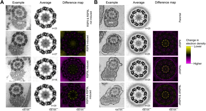
Fig. 5.
Both paralogues of RSP9 are necessary for RS head, but not overall axoneme, assembly in T. brucei and L. mexicana. (A) Ultrastructure change upon RSP9 and RSP9L RNAi in T. brucei. Transmission electron micrographs of transverse sections of detergent-extracted axonemes in induced TbRSP9RNAi, TbRSP9LRNAi and TbRSP9/9LRNAi cell lines in comparison to uninduced TbRSP9/9LRNAi cell lines. PFR, paraflagellar rod. (B) Ultrastructure change upon RSP9 and RSP9L deletion in L. mexicana. Electron micrographs of transverse sections of axonemes in ΔLmxRSP9, ΔLmxRSP9L and ΔLmxRSP9/9L in comparison to the parental cell line (Cas9T7). In A and B, the first column shows one representative axoneme cross-section. The second column shows an averaged axoneme structure, in which axoneme cross-sections have had perspective deviation from circularity corrected, followed by ninefold rotational averaging and averaging across multiple axoneme (n indicates the number of axonemes used). The third column shows an electron density difference map, resulting from subtraction of the induced RNAi or deletion mutant average axoneme image from the uninduced control or parental cell line. Yellow indicates more electron density in the uninduced control or parental cell line; magenta indicates more electron density in the induced RNAi or deletion mutant. There is a specific loss in electron density from the RS heads in the induced TbRSP9LRNAi, TbRSP9/9LRNAi, ΔLmxRSP9, ΔLmxRSP9L and ΔLmxRSP9/9L images.

