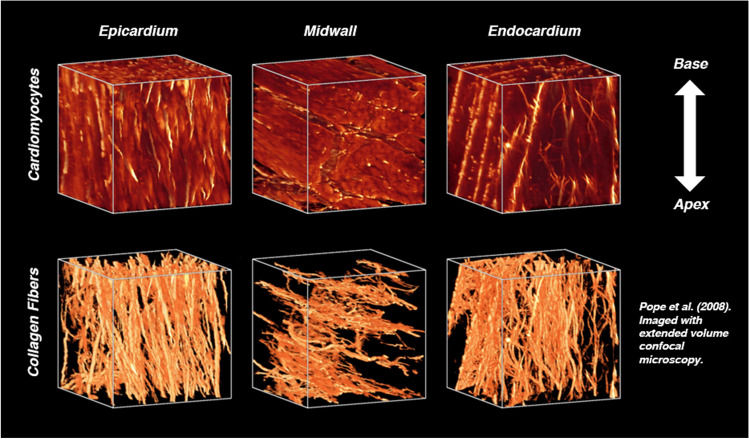
Fig. 1.
The orientations of cardiomyocytes and collagen fibers vary transmurally in the healthy rodent LV, but are co-oriented throughout the heart wall. The predominance of collagen fibers and their orientations, imaged here using extended volume confocal microscopy, give rise to nonlinear anisotropic mechanical behavior. Reprinted from the American Journal of Physiology—Heart and Circulatory Physiology, Vol. 295, A. Pope et al., “Three-dimensional transmural organization of perimysial collagen in the heart,” pp. H1243-1252. Copyright (2008), with paid permission from The American Physiological Society

