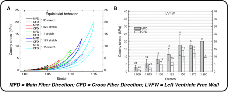
Fig. 2.
The microscopic structure and organization of myocardium’s healthy ECM gives rise to nonlinear force–displacement relationships, hysteresis, and anisotropic mechanical behavior with a preference for the circumferential (main fiber) direction during ex vivo equibiaxial testing. Each of these mechanical attributes can be seen in panel A (nonlinearity, hysteresis, anisotropy) and panel B (anisotropy, impressive passive extensibility), taken from equibiaxial extensions of human myocardium by Sommer et al. (2015). Reprinted from Acta Biomaterialia, Vol. 24, G. Sommer et al., “Biomechanical properties and microstructure of human ventricular myocardium,” pp. 172–192. Copyright (2015), with permission from Elsevier

