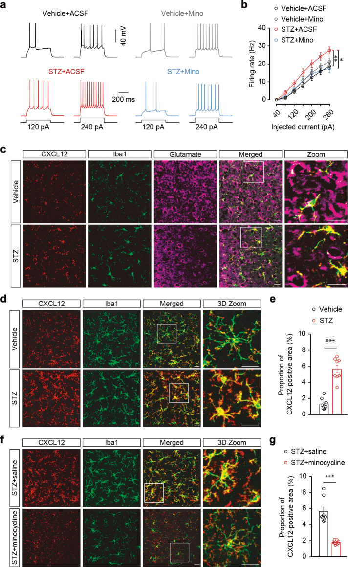
Fig. 4. The expression of microglial CXCL12 is increased in STZ 7W mice.
Representative traces (a) and summarized data (b) of action potentials recorded from glutamate neurons in the ACC slices from STZ- and vehicle-administered mice treated with ACSF or Mino (n = 22 neurons from three mice per group). Mino, minocycline. c Representative two-dimensional immunostaining images of CXCL12 (red), Iba1 (green), and glutamate (purple) in the ACC of STZ 7 W and vehicle mice. Scale bars, 20 μm. d Representative three-dimensional (3D) immunostaining images of CXCL12 (red) and Iba1 (green) in the ACC of STZ 7 W and vehicle mice. Three-dimensional reconstruction with ~30 two-dimensional immunostaining images. Scale bars, 20 μm. e Summarized data obtained by 3D image reconstruction showing the proportions of the CXCL12-positive area in the ACC of mice, as indicated in (d) (n = 8 slices from three mice per group). f Representative 3D immunostaining images of CXCL12 (red) and Iba1 (green) in the ACC of STZ 7 W mice treated for 2 weeks with minocycline or saline starting from the fifth week after STZ injection. Scale bars, 20 μm. g Summarized data showing the proportions of CXCL12-positive area in the ACC of mice, as indicated in (f) (n = 8 slices from three mice per group). Data are shown as means ± SEM. *P < 0.05; **P < 0.01; ***P < 0.001. Two-way repeated-measures ANOVA with Bonferroni post hoc analysis was used for (b). A two-tailed unpaired Student’s t test was used for (e, g)

