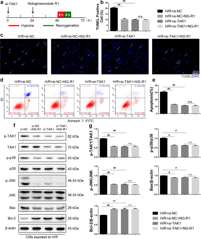
Fig. 7. NG-R1 inhibits H/R-induced apoptosis in CMs by suppressing TAK1.
a Schematic illustration of si-TAK1 transfection. CMs were transfected with si-TAK1 48 h before H/R injury. b, c Representative images and quantitative analysis of TUNEL-positive cells. Scale bar = 200 µm. d Representative images of apoptosis, as determined by FITC-PI/Annexin V staining. e Statistical analysis of apoptosis in the four groups. f The protein levels of p-TAK1, TAK1, p-p38, p38, p-JNK, JNK, Bcl-2 and Bax were determined by Western blotting. g Quantitative analysis of f. The data are shown as the mean ± SEM (n = 6 for each group). #P < 0.05 and ##P < 0.01 vs. the H/R + si-NC group.

