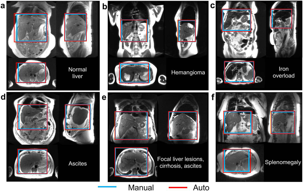
Figure 2.
3D detection of the liver in all three localizer orientations, with examples of common pathologies. a) In most cases, the liver volume was covered accurately by automated prescription. The automated prescription aligned well with the manual annotation in patients with focal lesions (b), iron overload (c), ascites (d), and/or cirrhosis (e), as well as in a patient with splenomegaly (f), where the spleen abuts the liver and its signal level is similar to that of liver, but the proposed method was still able to identify the liver correctly.

