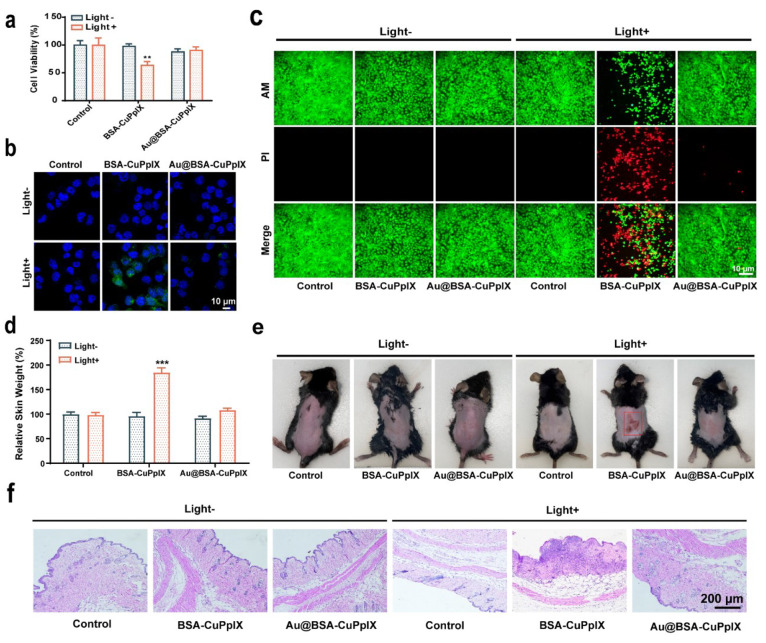
Figure 6.
Skin photoprotection evaluation. (a) The viability of HaCaT cells was assessed by CCK8 assay after being cultured with PBS, BSA-CuPpIX, and Au@BSA-CuPpIX, with or without LED light (405 nm) irradiation (5 W/cm2 for 5 min) at the same CuPpIX concentration. (b) The confocal imaging of HaCaT cells treated with PBS, BSA-CuPpIX, and Au@BSA-CuPpIX and then stained using DCFH-DA (green). The cell nuclei were stained using DAPI (blue). (c) The confocal imaging of live and dead cells stained by Calcein-AM and PI dyes. (d) Skin samples measuring 1.5 cm × 1.5 cm were collected to determine their relative weight after exposing the backs of euthanized mice to 405 nm LED light at a power density of 5 mW/cm2 for 2 h. (e) Photograph and (f) H&E sections of the skin tissue on the back of the mouse taken 3 days after with or without exposure to 405 nm LED light (5 mW/cm2, 2 h) treatments.

