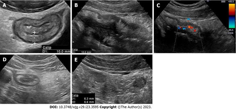
Figure 1.
Intestinal ultrasound assess several features of the bowel wall and surrounding tissue. A: Bowel wall thickness should be measured perpendicular to the anterior wall of the bowel (or where it is better visible) avoiding haustrations and mucosal folds. The cursor/calipers should be placed at the end of the interface echo between the serosa and the proper muscle to the start of the interface echo between the lumen and the mucosa; B: Bowel wall stratification. The wall layers in case of active disease may appear focally or extensively disrupted (disrupted or hypoechoic echo pattern); C: Bowel wall vascularity. Bowel wall vascularity can be determined both by colour Doppler or power Doppler signal at the level of the most thickened segments, using special presets optimized for slow flow detection; D: Mesenteric hypertrophy. Mesenteric hypertrophy also called fat wrapping or creeping fat appears on ultrasound (US) as hyperechoic tissue or “mass effect” encircling the diseased bowel; E: Lymphnodes. Enlarged inflammatory mesenteric lymph nodes related to Crohn’s disease are usually described at US as oval or elongated with lesser diameter > 5 mm and seem to be correlated with young age, early disease, or disease with shorter duration, and with the presence of fistulae and abscesses.

