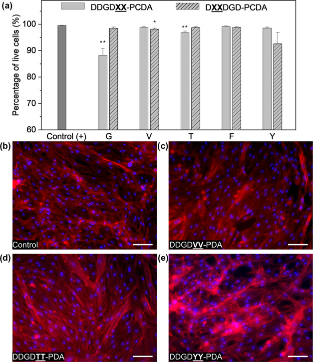
Figure 8.
Interfacing peptide-PDA monomers and their polymerized films with HDFs. (a) Cellular viability of HDFs incubated with 1 mM peptide-PDA after 6 h of exposure. Error bars represent the standard error of the mean; n = 6. For the statistical analysis, unpaired t-test with Welch’s correction was performed; *p < 0.05, **p < 0.01 vs positive control. Representative images of HDFs incubated for 5 days on films made from 5 mM peptide-PDA solutions at pH 2; HDFs on (b) glass coverslips and (c) 5 mM DDGDVV-PDA, (d) 5 mM DDGDTT-PDA, (e) 5 mM DDGDYY-PDA. Cells were stained with DAPI (nucleus) and Phalloidin (F-actin); Scale bar = 100 μm.

