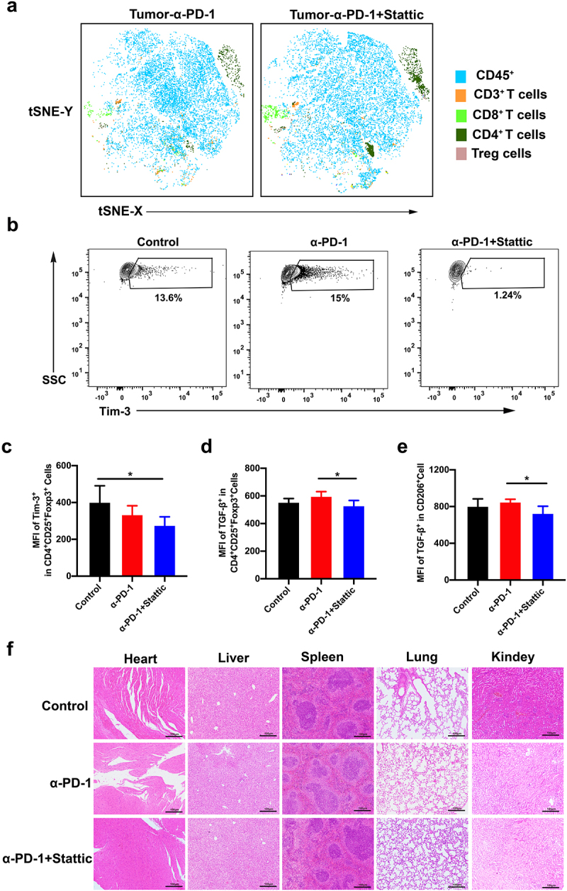
Figure 6.

Immune response of tumor microenvironment after combinatorial STAT3 inhibitor and PD-1 mAb therapy in the CT26 mouse model. (a) tSNE analysis for CD45+ T cells, CD3+ T cells, CD4+ T cells, CD8+ T cells and Treg cells in the tumor after combinatorial Stattic and PD-1 mAb therapy. (b-c) Represented contour images (b) and quantification of Tim-3 (c) in Treg cells after combinatorial Stattic and PD-1 mAb therapy in CRC tumor. (d-e) Quantification of MFI for TGF-β both in Tregs and M2 macrophages in CRC mouse model after different treatment. (f) No significant organ toxicity was assessed through H&E staining of samples of the heart, liver, spleen, lung, and kidneys in mouse after treatment.
