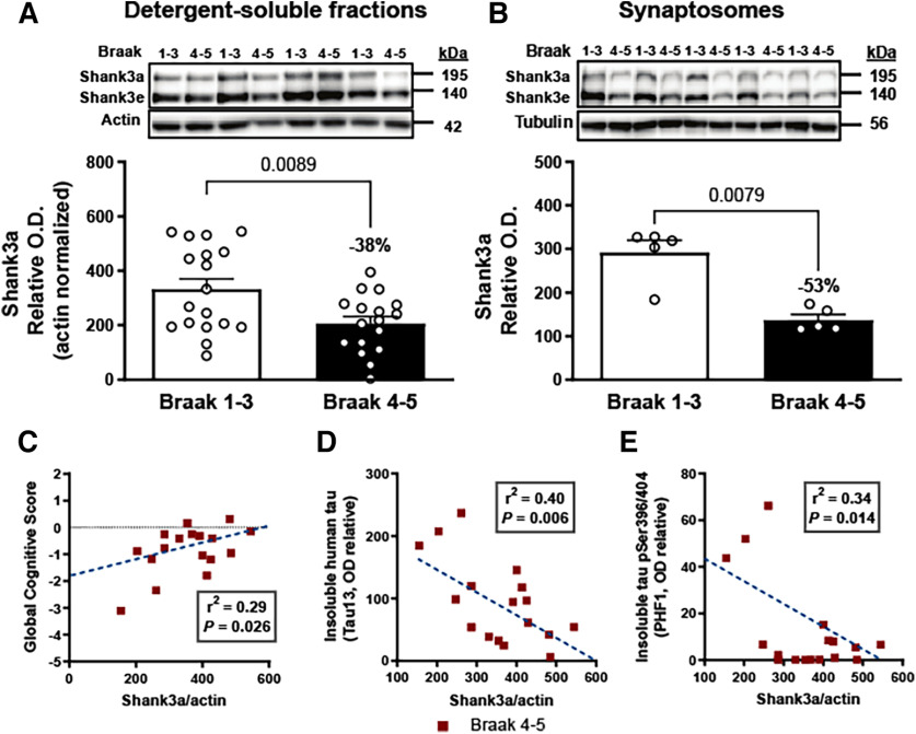
Figure 1.
Shank3a loss in AD correlates with disease severity. A, Shank3a (195 kDa) was quantified (antibody: ab136429) by WB in detergent-soluble extracts from parietal cortex of control (Braak Stages 1-3) and AD (Braak Stages 4 and 5) groups (n = 17 or 18). Shank3a levels were significantly reduced in individuals with AD. B, The difference in Shank3a levels between controls and AD subjects was more marked in synaptosome extracts (n = 5). Linear regression analyses showed that Shank3a levels in the parietal cortex correlated (C) positively with global cognitive scores and (D) negatively with levels of insoluble total tau (Tau13) and (E) Insoluble phospho-tau labeled with PHF1 antibody in persons with AD. Data are mean ± SEM. p < 0.05. A, B, Data were compared using the Mann–Whitney test. C-E, The coefficient of correlation r2 was calculated by linear regression. The p value was obtained using a generalized linear model.

