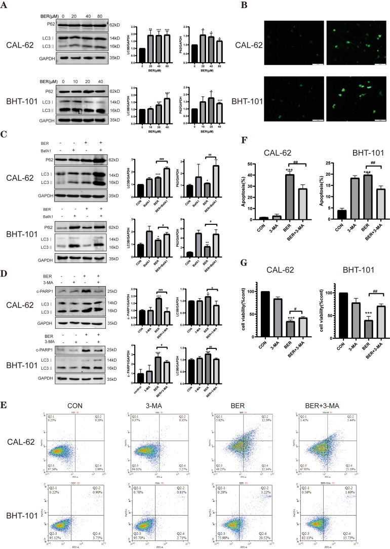
Figure 3.
BER-induced autophagy contributes to cell death in ATC cells. (A) The expression of autophagy-related proteins was evaluated in CAL-62 and BHT-101 cells after treatment with the indicated concentrations of BER for 72 h by Western blot analysis. GAPDH was used as a control. Representative column diagrams show the results of relative protein expression. (B) CAL-62 and BHT-101 cells were transiently transfected with GFP-LC3 expression plasmids for 12 h followed by treatment with 80 or 40 μM BER for 72 h. Scale bar, 100 μm. (C) ATC cells were pretreated with or without 2 μM BafA1for 2 h followed by treatment with or without BER for 72 h. Cells were then harvested for Western blot analysis to examine the LC3‑II/LC3‑I ratio (LC3B) and p62 levels. GAPDH was used as a control. Representative column diagrams showing the results of relative protein expression. (D) The cells were cultured with BER for 72 h in the absence or presence of 2 mM 3-MA, and Western blot analysis of the LC3‑II/LC3‑I ratio (LC3B) and c-PARP1 was performed. (E) Cells were treated as in D, and apoptosis was detected by flow cytometry. (F) The cell apoptosis rate was calculated from the flow cytometry results. (G) Cells were treated as in D, and the inhibitory rate was measured by CCK-8 assays. Data are expressed as the mean ± SD from three independent experiments. *P < 0.05, **P < 0.01, and ***P < 0.001 vs 0 µM BER or the control group; #P < 0.05, ##P < 0.01, and ###P < 0.001 vs the BER group.

