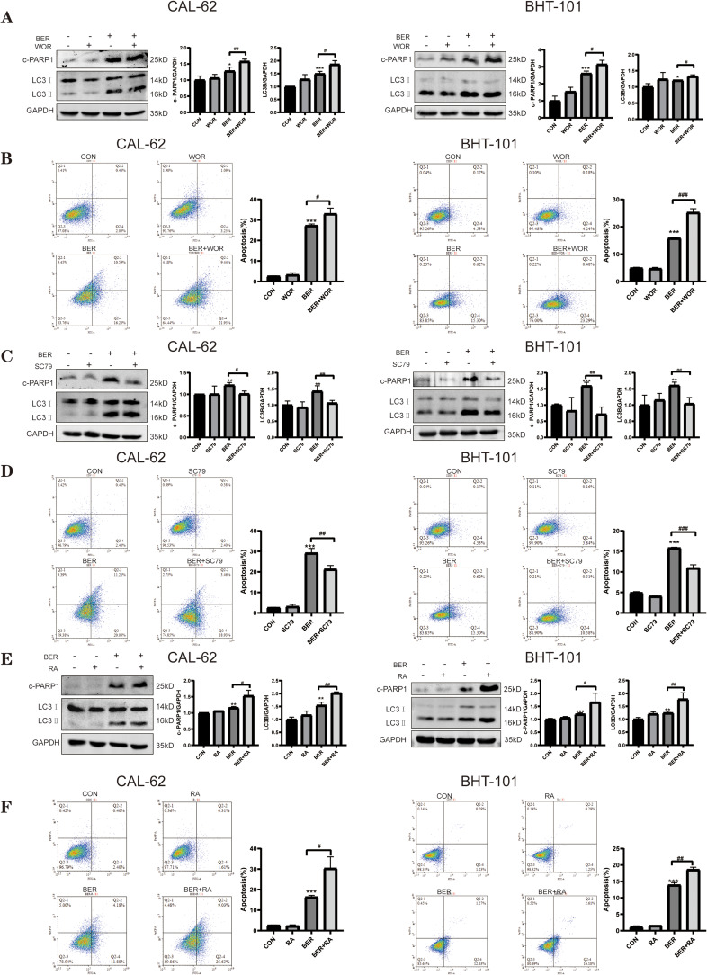
Figure 5.
BER mediates autophagy and apoptosis by regulating the PI3K/AKT/mTOR signaling pathway. CAL-62 and BHT-101 cells were pretreated with 1 μMWOR (a PI3K inhibitor), 4 μg/mlSC79 (an AKT activator), and 0.2 μM RA (anmTOR inhibitor) for 2 h prior to 80 or 40 μM BBR treatment for 72 h. (A) Cells were treated with or without BER in combination with or without 1 μM WOR, and the levels of the apoptosis-related protein, cleaved PARP1, and the autophagy-related protein, LC3B, were determined by Western blot analysis. (B) Cells were treated as in A, and apoptosis was detected using flow cytometry. (C) Cells were treated with or without BER in combination with or without 4 μg/mL SC79, and the levels of the apoptosis-related protein, cleaved PARP1, and the autophagy-related protein, LC3B, were determined by Western blot analysis. (D) Cells were treated as in C, and apoptosis was detected using flow cytometry. (E) Cells were treated with or without BER in combination with or without 0.2 μM RA, and the levels of the apoptosis-related protein, cleaved PARP1, and the autophagy-related protein, LC3B, were determined by Western blot analysis. (F) Cells were treated as in E, and apoptosis was detected using flow cytometry analysis. Data are expressed as the mean ± SD of triplicate experiments. *P < 0.05, **P < 0.01, and ***P < 0.001 vs the control group; #P < 0.05, ##P < 0.01, and ###P < 0.001 vs the BER group.

