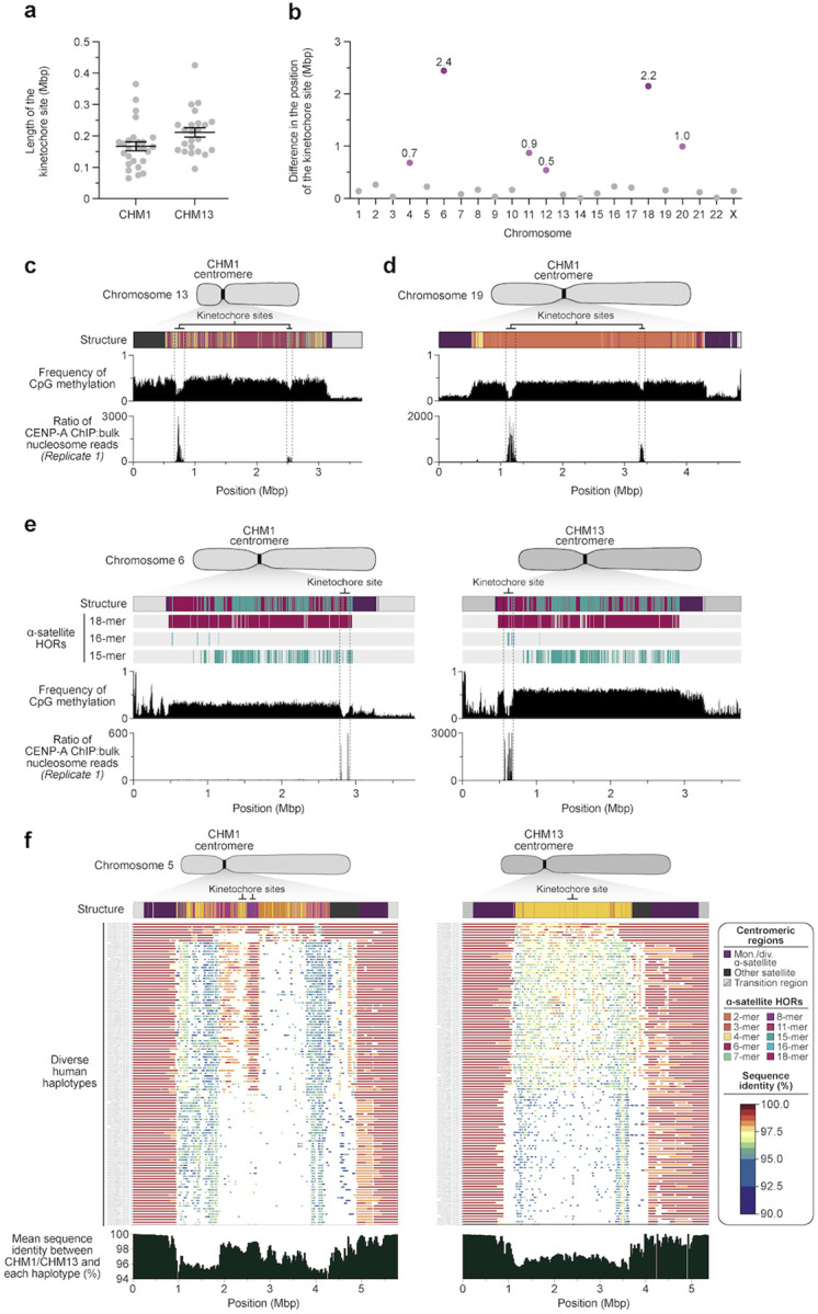
Figure 4. Variation in the site of the kinetochore among two sets of human centromeres.

a) Plot comparing the length of the kinetochore site, marked by hypomethylated DNA and CENP-A-containing chromatin, between the CHM1 and CHM13 centromeres. b) Plot showing the difference in the position of the kinetochore among the CHM1 and CHM13 centromeres. c,d) Discovery of two potential kinetochores on the c) chromosome 13 and d) chromosome 19 centromeres in the CHM1 genomes. The presence of two hypomethylated regions enriched with CENP-A chromatin likely represents two populations of cells, which may have arisen due to a somatic mutation, resulting in differing epigenetic landscapes. e) Comparison of the CHM1 and CHM13 chromosome 6 centromeres, which differ in kinetochore position by 2.4 Mbp. f) Comparison of the CHM1 and CHM13 chromosome 5 centromeres, showing that the sequences underlying the CHM1 kinetochore are conserved in approximately half of the HPRC genomes, but the same degree of conservation is not observed for the CHM13 kinetochore region.
