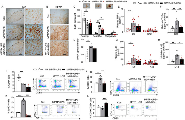
Figure 3. Systemic NDP-MSH treatment reduces neuroinflammation and modulates peripheral immune responses.
C57BL/6 mice were treated i.p. with MPTP.HCl (20 mg/kg) and LPS (1 mg/kg) from day (D) 1 to D4 and NDP-MSH (400 μg/kg) or vehicle control from D1 to D12. Plasma was collected at D5 and D12. Mice were sacrificed at D12. (A) Cells stained positive for iba1 in the SN. Scale bars, 100 μm and 30 μm. (B) GFAP staining. Scale bar, 30 μm. (C) Morphological classification and quantification of iba1+ microglia. Two-way ANOVA followed by Tukey’s post hoc test; n=5/group. (D) Quantification of integrated optical density of GFAP in the SN. One-way ANOVA followed by Tukey’s post hoc test; n=3/group. ELISA assessment of TNF-α levels in (E) plasma at D5 and D12 and (F) ventral midbrain at D12 and IL-1β in (G) plasma at D5 and D12 and (H) ventral midbrain at D12. Two-way ANOVA followed by Tukey’s post hoc test. **p<0.01; n=5–7/group/time point. Flow cytometric analysis of the splenocytes showing percentages of (I) CD4+ helper T cell, (J) CD8+ cytotoxic T cells, (K) LY6C+ cell, and (L) CD4+CD25+ Tregs. One-way ANOVA followed by Tukey’s post hoc test; *p<0.05, **p<0.01; ***p<0.001; n=4–5/group.

