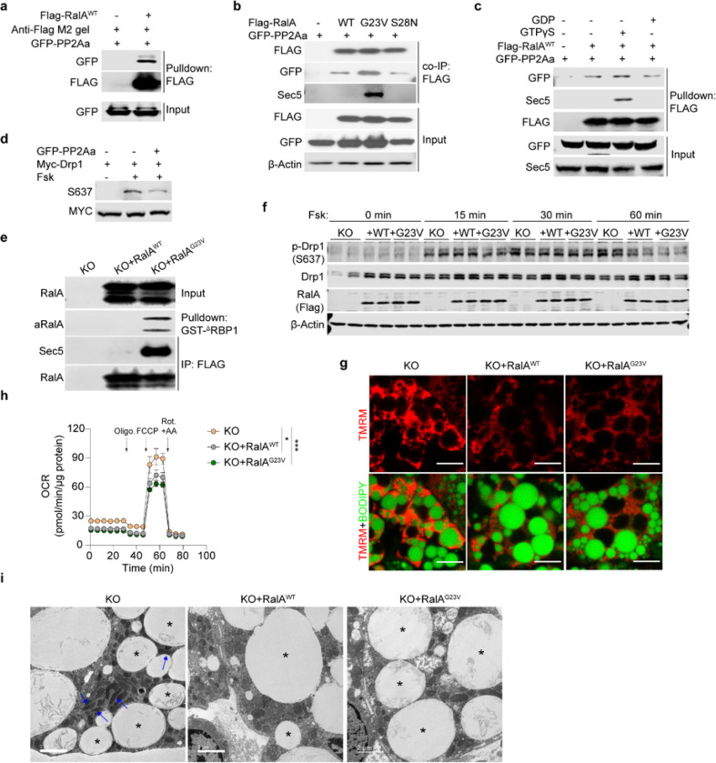
Figure 6. RalA interacts with Drp1 and protein phosphatase 2A, promoting dephosphorylation of Drp1 at S637.
a, Pull down assay determining PP2Aa-RalA interaction. Purified Flag-RalAWT was used to pull down GFP-PP2Aa overexpressed in HEK293T cells, b, Immunoblot analysis of co-immunoprecipitation determining interaction between RalA wildtype (WT), constitutive active (G23V), or dominant negative (S28N) mutants and PP2Aa in HEK293T cells, c, Flag-pull down assay determining interaction between PP2Aa and GTP/GDP-loaded RalA. Purified Flag-RalAWT protein loaded with either GTPγS or GDP was respectively used as a prey to pull down GFP-PP2Aa from HEK293T cells, d, In vitro dephosphorylation assay in HEK293T cells co-transfected with PP2A and Drp1 plasmids. Cells were treated for 1 hr with 20 μM forskolin (Fsk) or vehicle, e, RalA activity assay in immortalized RalA KO adipocytes reconstituted with RalAWT and RalAG23V. f, Immunoblottmg of phospho-Drp1 (S637), total Drp1, Flag-tagged RalA and β-Actin in immortalized RalA KO adipocytes with or without RalA reconstitution (n = 3). Adipocytes were treated with 20 μM forskolin for indicated time, g, Representative confocal images of live immortalized adipocytes (n = 3) stained with TMRM (red) and BODIPY (green), scale bar = 15 μM. h, OCR was measured by seahorse in immortalized adipocytes (n = 5–6). Vertical arrows indicate injection ports of indicated chemicals. Data are shown as the mean ± SEM, *p < 0.05, ***p < 0.001 by two-way ANOVA. i, Representative EM images (n = 3) of RalA KO immortalized adipocytes with or without RalA reconstitution. Blue arrow indicates elongated mitochondria; asterisk indicates lipid droplet. Scale bar = 2 μm.

