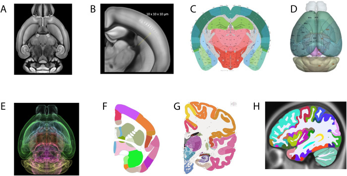
Fig 3. Common coordinate frameworks of the brain.
(A, B) Allen Mouse Brain Common Coordinate Framework (CCF) constructed from serial 2-photon tomography images with 100 μm z-sampling from 1,675 young adult C57BL/6J mice yields 10-μm cubic resolution. (C) Digital atlases of the Mouse (Allen CCFv3) annotated plate and (D) 3D reconstruction. (E) fMOST mouse atlas derived from CCFv3 through iterative averaging of 36 fMOST brains. This approach to a reference atlas reduces the average distance error of somata mapping up to 40% (F) marmoset atlas plate (Allen Institute for Brain Science). (G) Human reference atlas from 34-year-old female, 1 mm/pixel Nissl and immunohistochemistry anatomical plates, annotated 862 structures, including 117 white matter tracts and several novel cyto- and chemoarchitecturally defined structures. (H) MRI-based annotation of human atlas of 150 structures form the initial atlas for BICAN profiling.

