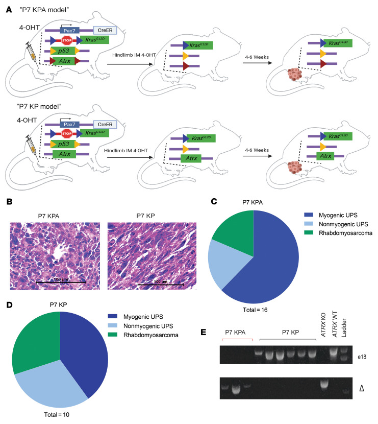
Figure 2. Primary mouse model of Atrx-deleted soft tissue sarcoma.
(A) Schematic showing the spatially and temporally restricted primary mouse model of Atrx-deleted soft tissue sarcoma. 4-hydroxytamoxifen (4-OHT) is injected into the gastrocnemius muscle, which leads to activation of the CreER expressed from the endogenous Pax7 promoter in muscle satellite cells to activate expression of KrasG12D and delete floxed Trp53 and Atrx alleles. Atrx is deleted in P7 KPA mice (top) and a WT Atrx is retained in control P7 KP mice (bottom). Loxp sites are designated by colored triangles in the diagram. (B) H&E staining of sarcomas from P7 KPA and P7 KP mice. These H&E images are also shown in Supplemental Figure 2A (left) accompanied by staining for myogenic markers. (C and D) Classification of tumor type for P7 KPA (n = 16) and P7 KP (n = 10) tumors, as determined by IHC for 9 myogenic and other markers. Myogenic UPS was defined as when cells had pleomorphic nuclei characteristic of UPS but stained positive for at least 2 of the 4 tested myogenic markers (MyoD1, Myogenin, Desmin, and SMA). (E) Genotyping assays to confirm complete deletion of Atrx in the P7 KPA tumor model. Genotyping of sarcoma cell lines from P7 KP and P7 KPA mouse tumors for the presence of loxP flanked Atrx exon 18. The top genotyping gel portion shows the presence or absence of the portion of the Atrx band targeted for excision by the Cre/loxP system, with absence of a band in E1 indicating successful deletion of exon 18 of Atrx. To confirm the findings in the top gel, an additional genotyping assay (bottom) was performed that selectively amplified only the sequence that occurs after successful deletion of exon 18. The presence of a band in the bottom gel indicates successful deletion of Atrx exon 18.

