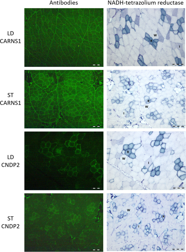
Fig. 2.
Immunohistochemical detection of carnosine synthase (CARNS1) and carnosine dipeptidase 2 (CNDP2) enzymes in serial cross sections of the longissimus dorsi (LD) and semitendinosus (ST) muscles. Serial sections were stained with anti-CARNS1 and anti-CNDP2 antibodies (left panels, green staining) or with a histochemical reaction based on NADH-tetrazolium reductase activity (right panels) to identify white glycolytic (W), intermediate (I) and red oxidative (R) muscle fibres. CARNS1 staining is mainly found at the periphery of W and R muscle fibres. A strong CNDP2 signal is also observed at the periphery of muscle fibres, with stronger intensity found in red oxidative fibres. Diffuse cytosolic CARNS1 and CNDP2 staining is also present in all fibre types, with slightly stronger intensity found in red oxidative fibres. Scale bar = 200 µm

