Fig. 2.
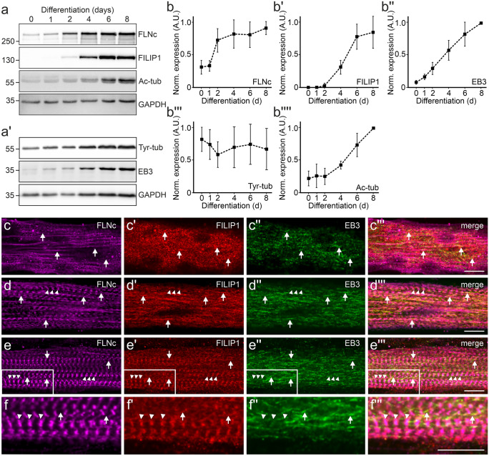
FILIP1 expression and localization in differentiating cultured C2C12 myotubes. a, a' Representative western blots showing the levels of FLNc, FILIP1, EB3, tyrosylated tubulin (Tyr-tub) and acetylated tubulin (Ac-tub) in proliferating C2C12 myoblasts (0 days) and myotubes differentiated for the indicated time (1–8 days). GAPDH was used as loading control and for normalization. b–b'''' Quantification of the data presented in (a–a'), expressed as mean ± SD. n = 3. In all individual experiments maximum relative expression of each protein was set to 1.0. c–e''' Immunolocalization of FLNc, FILIP1 and EB3 in C2C12 myotubes differentiated for 2 (c–c'''), 4 (d–d''') and 6 (e–e''') days. c–c''' FILIP1 and EB3 partially colocalize (arrows), which becomes more evident later in differentiation (d–d''', arrows). This is still evident in mature myotubes 6 days after the start of differentiation (e–e''', arrows), when FILIP1 and FLNc are mainly present in Z-discs (e–e''' and f–f''', arrowheads). A magnification of the boxed area in (e–e''') is shown in (f–f'''). Bars: 10 μm

