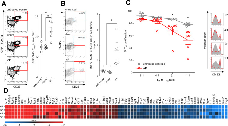
Figure 4.
Immune suppressive function of Tregs during AP. AP was induced by partial duct ligation in DEREG- and C57Bl/6 mice. (A) The number of splenic GFP producing Tregs was increased in the AP group (n=5) but not in control or sham operated mice (n=3). (B) The same increase of FOXP3+/CD25+/CD4+ Tregs was observed in the small intestine. (n=5). (C) Suppression assays showed that Tregs from AP mice (red line) have an increased suppressive capacity on Teff cell proliferation. (D) Heat map illustrates fold changes of gene transcription in Tregs from AP mice (n=4) compared with untreated controls (n=4). Statistically significant differences were tested by unpaired Student’s t-test for independent samples and significance levels of p<0.05 are marked by an asterisk, corrected for multiple testing (bonferroni-correction). AP, acute pancreatitis.

