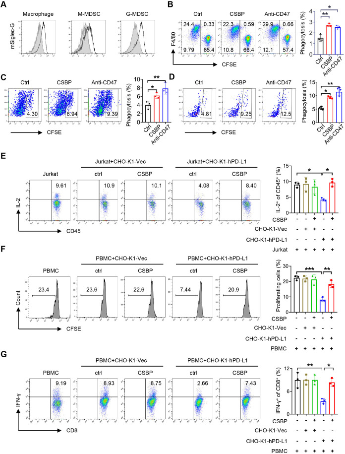
Figure 3.
4T1 cancer cells phagocytosis by macrophage/M-MDSC and T-cell activation after CSBP treatment. (A) Representative flow cytometry histogram of the expression of mSiglec-G (black lines) versus isotype control (gray shaded curves) by macrophage, M-MDSC or G-MDSC. (B) Phagocytosis assays were performed with CD24+ 4T1 cells by BMDMs in the presence of CSBP or anti-CD47 mAb. (C, D) Flow cytometry analysis of phagocytosis of 4T1 cells by macrophages and M-MDSCs from 4T1 tumor-bearing mice spleen, in the presence of CSBP or anti-CD47 mAb. (E) Evaluation of the effects of CSBP on IL-2-expressing Jurkat T cells. Intracellular staining of IL-2 was detected by flow cytometry. (F, G) PBMCs from healthy donors were isolated and stained with 0.2 µM CFSE. Then, PBMCs were activated with 100 U/mL IL-2, 1 µg/mL anti-CD3, and 1 µg/mL anti-CD28 stimulatory antibodies, and cultured with CHO-K1-Vec or CHO-K1-hPD-L1 cells with or without CSBP. After 3 days, the proliferation of CD8+ T cells and proportion of IFN-γ-expressing CD8+ T cells were analyzed by flow cytometry. Data are representative of at least three independent experiments. Data are presented as means±SEM, and statistical significance was determined by two-way analysis of variance with multiple comparisons. *p<0.05, **p<0.01, ***p<0.001. BMDMs, bone marrow-derived macrophages; CFSE, carboxyfluorescein succinimidyl ester; CSBP, CD24/Siglec-10 blocking peptide; G-MDSC, granulocytic myeloid-derived suppressor cell; IFN, interferon; IL, interleukin; mAb, monoclonal antibody; M-MDSC, monocytic myeloid-derived suppressor cell; PBMCs, peripheral blood mononuclear cells.

