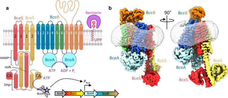
Fig. 1. Organization of the BceAB-S complex.
a Diagram showing the topology and stoichiometry of the BceAB-S membrane protein complex. The transmembrane (TM) helices of BceB are colored blue, green, and orange, and labeled with white numbers. BceS monomers are colored red and yellow with TM helices indicated with black numbers. Individual domains in the cytoplasmic region of BceS are labeled in black. Bacitracin binding to the lipid target UPP is shown as a pink crescent surrounding the phosphates of UPP. b Cryo-EM map of the BceAB-S membrane protein complex in a nucleotide-free state. Protein components are colored the same as in a. The detergent micelle is shown in grey surrounding the TM helices.

