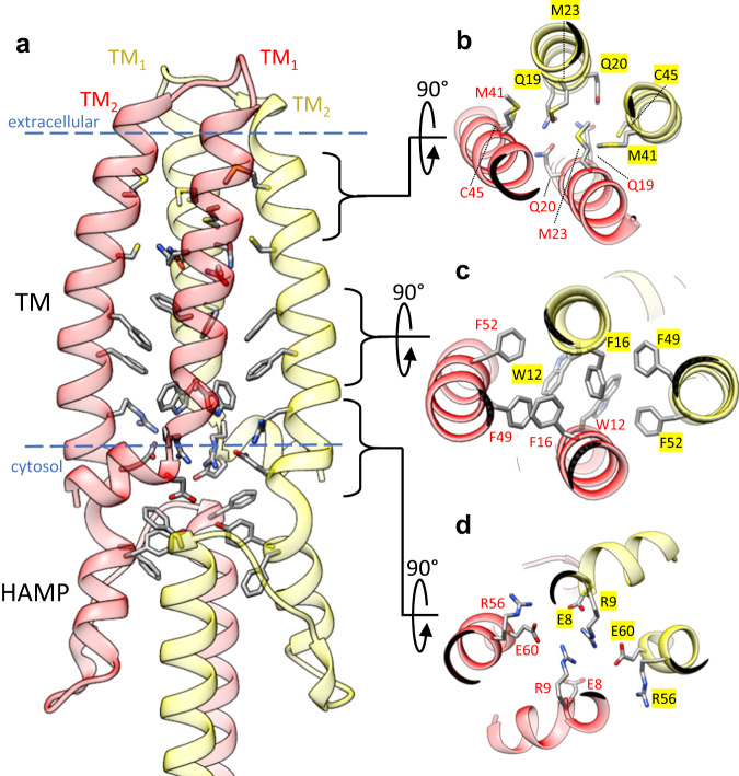
Fig. 2. BceS TM helix interactions.
a Overall configuration of the BceS TM helices and HAMP domain. Each monomer of BceS is colored either red or yellow. Blue dashed lines indicate the approximate boundary of the lipid bilayer. b View from the extracellular space of the tip of BceS TM helices. The core of the TM helix bundle contains a cluster of sulfur containing side-chains and hydrogen bonds between two pairs of glutamines. c View from the extracellular space of the middle of the BceS TM helix bundle, showing that the core of the bundle in the membrane interior contains a network of π-stacking interactions between aromatic side-chains. d View of the BceS TM helix bundle near the interface of the membrane and cytosol. An electrostatic network of charged residues mediates interaction between BceS TM helices.

