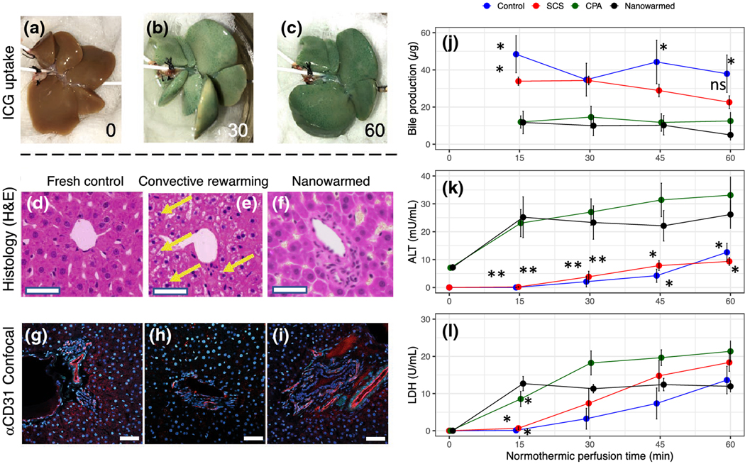
Figure 5. Organ-level function in vitrified and nanowarmed livers.
Vitrified and nanowarmed livers were tested for function and injury as compared to static cold storage (SCS, 18–20 hr in UW solution at 4 °C), CPA loaded and unloaded but not vitrified and untreated (flushed with UW and used within 30 min) controls. (A-C) Following washout of CPA+sIONP, the organs were assessed by normothermic machine perfusion with indocyanine green (ICG) in the perfusate and imaged at 0, 30, and 60 minutes. The nanowarmed livers demonstrated organ-level function with homogenous ICG uptake. (D-F) Histologic examination (hematoxylin and eosin) of control (left), vitrified and convectively rewarmed (middle), and vitrified and nanowarmed (right) livers show preserved tissue architecture in nanowarmed livers. Convectively rewarmed livers had diffuse areas of extra- and intracellular white space (yellow arrows) suggestive of ice crystallization during rewarming or cytoplasmic vacuolization. Nanowarmed livers have a normal portal and sinusoidal architecture. Bar = 100 μm. (G-I) Confocal imaging with anti-CD31 immunofluorescence shows that control and nanowarmed livers have preserved vascular architecture (red staining), whereas convectively rewarmed livers have reduced staining intensity, indicating endothelial injury. (J) Bile production assessed during 60 minutes of normothermic perfusion after nanowarming was not different than CPA-loaded and unloaded livers. Hepatocyte injury was assessed by measuring ALT (K) and LDH (L) in the SHVC venous effluent during perfusion. ALT levels for nanowarmed livers were slightly higher than for control organs throughout the normothermic perfusion, whereas LDH levels were not different between nanowarmed livers and other groups after 15 minutes. For all groups, n = 3–5. P values show comparison to nanowarming only with * = P <0.05 and ** = P <0.01. All significant differences are shown. Full pairwise comparison statistics are in the Supplementary Materials.

