
Fig. 2.
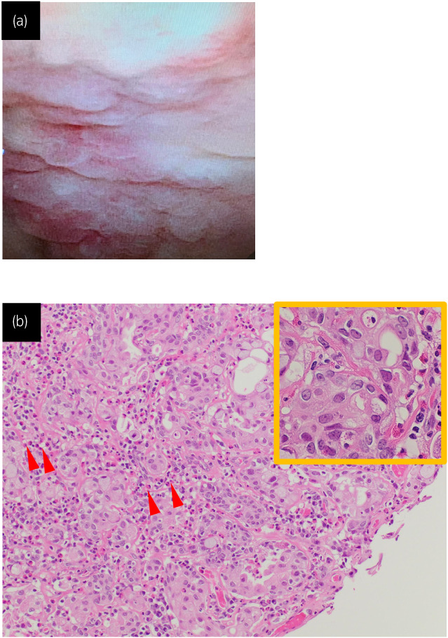
Cystoscopy revealed an erythematous mucosal lesion all over the bladder wall (a). Histopathologic examination of the specimen obtained by transurethral resection showed pT1 urothelial carcinoma and moderate infiltration of eosinophils (arrow head). (b). The tumor cells showed severe nuclear atypia on the high magnification. Hematoxylin and eosin (H&E) staining. Although a portion of the muscle layer was sampled, the tumor cells were found to only invade the subepithelial connective tissue, with no intramuscular invasion detected.