Figure 2.
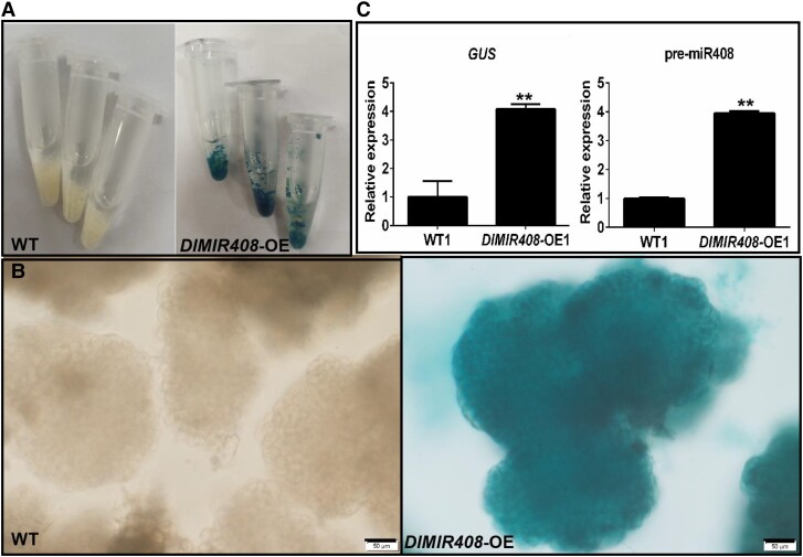
The expression pattern of GUS and the pre-miR408, and the cell morphology in WT and DlMIR408-OE cell line for the whole-transcriptome sequencing. A) The WT and DlMIR408-OE cell lines after GUS staining, and the cell lines with independent 3 biological replicates. B) The positive homozygous transgenic GE of DlMIR408-OE under Olympus microscope observation, scale bars = 50 μm. C) The expression patterns of GUS and pre-miR408 in WT and DlMIR408-OE1. Error bars indicate means ± SDs, n = 3. **Highly significant difference (Duncan’s post hoc test, P ≤ 0.01).

