. 2023 Apr 25;307(5):e223128. doi: 10.1148/radiol.223128

Figure 2:

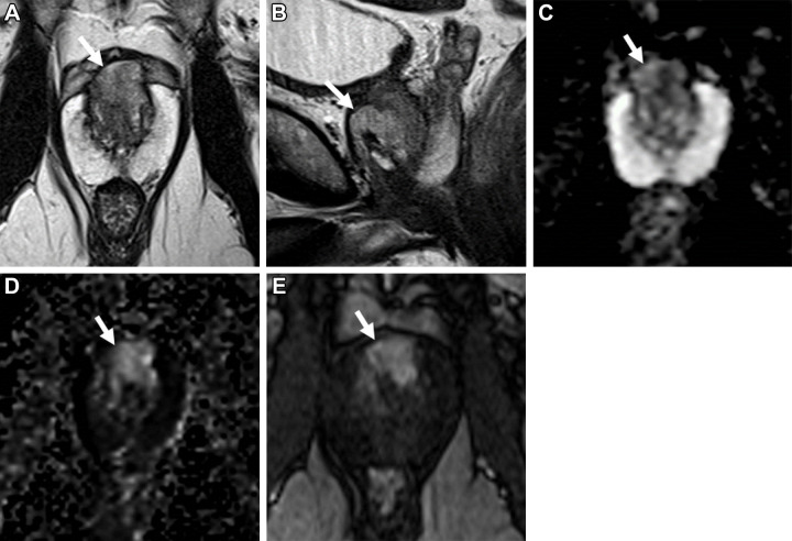
Images in a 68-year-old man with a prostate-specific antigen level of 21.8 ng/mL and a prior negative prostate biopsy. (A) Axial and (B) sagittal T2-weighted MRI scans show a lesion in the midline anterior transition zone at the mid gland, with intermediate to high signal intensity and anterior extraprostatic extension (arrows). (C) The apparent diffusion coefficient map shows diffusion restriction with moderately hypointense signal (arrow) within the lesion, while the (D) diffusion-weighted image with a high b value of 1400 sec/mm2 shows moderately hyperintense signal (arrow); the (E) dynamic contrast-enhanced MRI scan shows corresponding early arterial enhancement (arrow). The signal intensity of the lesion at T2-weighted MRI is higher than expected for typical prostate adenocarcinoma, but because of the extraprostatic extension findings, the lesion was assigned a T2-weighted MRI and an overall Prostate Imaging Reporting and Data System score of 5. MRI-targeted biopsy of the lesion revealed Gleason 4+4 prostate cancer with predominate cribriform morphology.

Images in a 68-year-old man with a prostate-specific antigen level of 21.8 ng/mL and a prior negative prostate biopsy. (A) Axial and (B) sagittal T2-weighted MRI scans show a lesion in the midline anterior transition zone at the mid gland, with intermediate to high signal intensity and anterior extraprostatic extension (arrows). (C) The apparent diffusion coefficient map shows diffusion restriction with moderately hypointense signal (arrow) within the lesion, while the (D) diffusion-weighted image with a high b value of 1400 sec/mm2 shows moderately hyperintense signal (arrow); the (E) dynamic contrast-enhanced MRI scan shows corresponding early arterial enhancement (arrow). The signal intensity of the lesion at T2-weighted MRI is higher than expected for typical prostate adenocarcinoma, but because of the extraprostatic extension findings, the lesion was assigned a T2-weighted MRI and an overall Prostate Imaging Reporting and Data System score of 5. MRI-targeted biopsy of the lesion revealed Gleason 4+4 prostate cancer with predominate cribriform morphology.