. 2023 Apr 25;307(5):e223128. doi: 10.1148/radiol.223128

Figure 3:

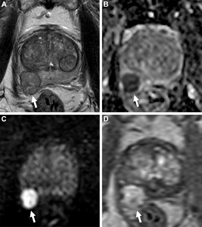
Images in a 78-year-old man with a serum prostate-specific antigen level of 10 ng/mL. (A) Axial T2-weighted MRI scan shows a well-encapsulated nodule in the right mid peripheral zone (arrow), which suggests an ectopic benign prostatic hyperplasia nodule. (B) The apparent diffusion coefficient map and (C) diffusion-weighted image with a b value of 1500 sec/mm2 show the nodule with diffusion restriction with prominent hypointense and hyperintense signal features (arrows), and the (D) dynamic contrast-enhanced (DCE) MRI scan shows focal early enhancement (arrow). The T2-weighted imaging, diffusion-weighted imaging, DCE MRI, and overall Prostate Imaging Reporting and Data System scores of this lesion were 2, 5, positive, and 5, respectively. Transrectal US/MRI–fusion guided biopsy revealed Gleason 4+4 prostate cancer within this lesion.

Images in a 78-year-old man with a serum prostate-specific antigen level of 10 ng/mL. (A) Axial T2-weighted MRI scan shows a well-encapsulated nodule in the right mid peripheral zone (arrow), which suggests an ectopic benign prostatic hyperplasia nodule. (B) The apparent diffusion coefficient map and (C) diffusion-weighted image with a b value of 1500 sec/mm2 show the nodule with diffusion restriction with prominent hypointense and hyperintense signal features (arrows), and the (D) dynamic contrast-enhanced (DCE) MRI scan shows focal early enhancement (arrow). The T2-weighted imaging, diffusion-weighted imaging, DCE MRI, and overall Prostate Imaging Reporting and Data System scores of this lesion were 2, 5, positive, and 5, respectively. Transrectal US/MRI–fusion guided biopsy revealed Gleason 4+4 prostate cancer within this lesion.