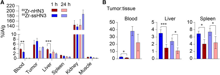
FIGURE 4.
Single-domain antibody PET tracers successfully image GPC3+ liver tumor xenografts. Shown are selected ex vivo biodistribution of 89Zr-ssHN3 and 89Zr-nHN3 (A) and tumor-to-tissue ratios of HepG2 tumor-bearing mice (n = 4) (B). Full 12-organ biodistribution results for mice bearing HepG2 tumors are reported in Supplemental Figures 19 and 20. *P < 0.05. **P < 0.01. ***P < 0.005.

