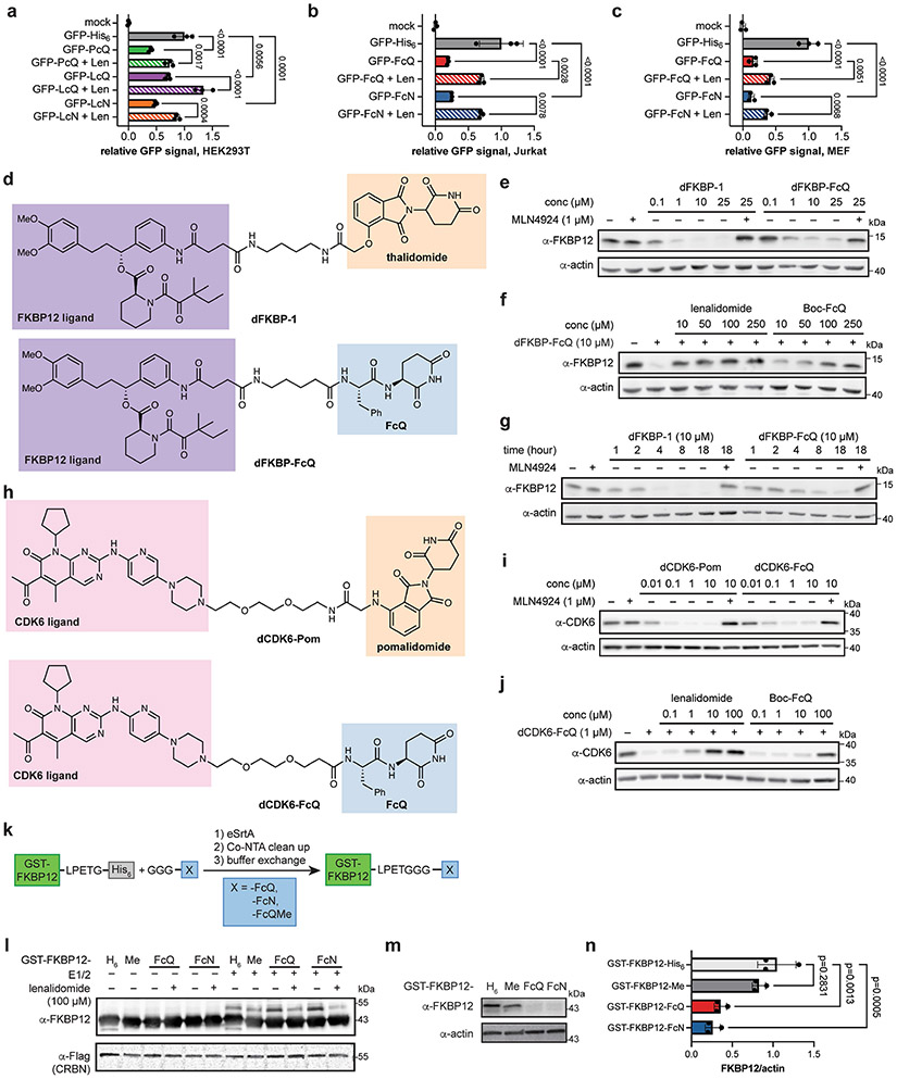
Extended Data Fig. 6 ∣. The C-terminal cyclic imide degron is transferrable.
(a-c) Flow cytometry analysis of the GFP levels in (a) HEK293T cells, (b) Jurkat or (c) MEF cells 6 h after electroporation with GFP tagged with the indicated peptide, with or without lenalidomide competition (100 μM). Comparisons were performed using an ordinary one-way ANOVA with Šídák’s multiple comparisons test, and p values are shown above comparison bars. (d) Structure of FKBP12 degraders dFKBP-1 and dFKBP-FcQ. (e) Western blot of FKBP12 levels after treatment of HEK293T cells with dFKBP-1 or dFKBP-FcQ over a 0.1–25 μM dose-response range. (f) Western blot of FKBP12 levels after co-treatment of HEK293T cells with dFKBP-FcQ and lenalidomide or Boc-FcQ. (g) Levels of FKBP12 over time in HEK293T cells treated with dFKBP-1 or dFKBP-FcQ. (h) Structure of CDK6 degraders dCDK6-Pom and dCDK6-FcQ. (i) Western blot of CDK4/6 levels after treatment of Jurkat cells with dCDK6-Pom or dCDK6-FcQ over a 0.01–10 μM dose-response range. (j) Western blot of CDK6 levels after co-treatment of Jurkat cells with dCDK6-FcQ and lenalidomide or Boc-FcQ. (k) Sortase system used to generate degron-tagged GST-FKBP12 from GST-FKBP12-LPETG-His6. (l) In vitro ubiquitination of FKBP12 tagged with C-terminal cyclic imide. FKBP12-H6 = FKBP12 with C-terminal His6 tag (no sortase treatment); FKBP12-Me = FKBP12 with C-terminal FcQMe; FKBP12-FcQ = FKBP12 with C-terminal FcQ; FKBP12-FcN = FKBP12 with C-terminal FcN. (m) Western blot of FKBP12 tagged with the indicated peptides 6 h after electroporation into HEK293T cells. (n) Quantification of Western blot in (m). Error bars represent mean ± SD. Comparisons were performed using an ordinary one-way ANOVA with Šídák’s multiple comparisons test. All western blot data are representative of at least 2 independent replicates. Flow cytometry data is representative of 3 independent replicates. For uncropped western blot images, see Supplementary Fig. 10.

