Figure 5.
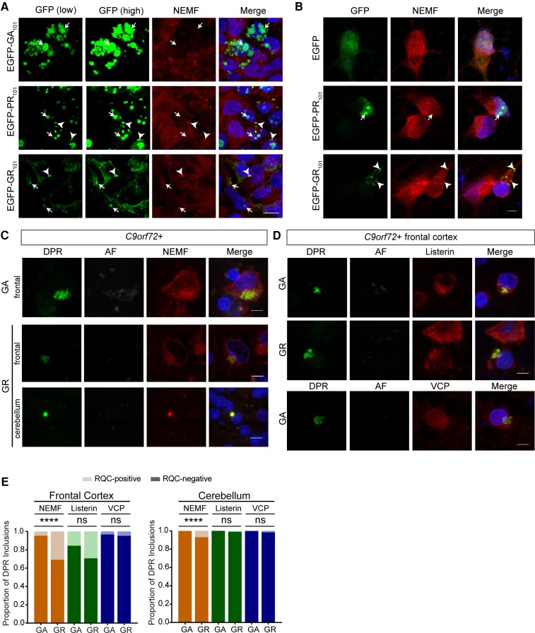
NEMF is recruited to DPR inclusions. (A) Representative confocal images of colocalization of NEMF and R-rich DPR proteins (arrowheads) expressed in HEK 293T cells (scale bar = 10 µm). (B) Representative confocal images of colocalization of NEMF and R-rich EGFP-DPR reporters (arrowheads) in I3N neurons (scale bar = 5 µm). (C) Confocal images show colocalization of DPR inclusions (poly-GA and poly-GR) and NEMF in frontal cortex and cerebellar granular layer of C9orf72 human tissue (scale bar, 5 µm). (D) Representative double immunofluorescence confocal images of DPR inclusions (poly-GA and poly-GR) and RQC factors (listerin and VCP) in frontal cortex of C9orf72-mutation carriers (scale bar = 5 µm). (E) Quantification of proportion of DPR inclusions (poly-GA and poly-GR) that co-localize with RQC complex factors NEMF, listerin and VCP in frontal cortex and cerebellum from five C9orf72-expansion cases (total number of inclusions counted for poly-GA n = 90–130 or n = 1049–1201 and for poly-GR n = 41–55 or n = 62–114 in frontal cortex and cerebellum, respectively; frontal cortex: Fisher’s exact test, NEMF P < 0.0001, listerin P = 0.0668 and VCP P = 0.6507; cerebellum: Fisher’s exact test, NEMF P < 0.0001, listerin P = 0.0767 and VCP P = 0.1403).

