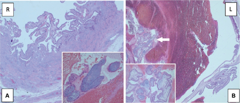
Figure 3.

(a, b) Right fallopian tube: Edema and decidualized stromal areas in the perforated tubal wall (H&E 25x). Degenerated villi structures in the blood-fibrin mass falling from the perforated area of the fallopian tube (H&E 200x) b. Left fallopian tube: The villi structures between the blood-fibrin masses (shown by the arrow) in the lumen of the fallopian tube (H&E 25x). Magnification of villus structures (H&E 200x).
