
Figure 5: ADAR1 negatively regulates Drosha-DGCR8 interaction in macrophages and AAA lesions.
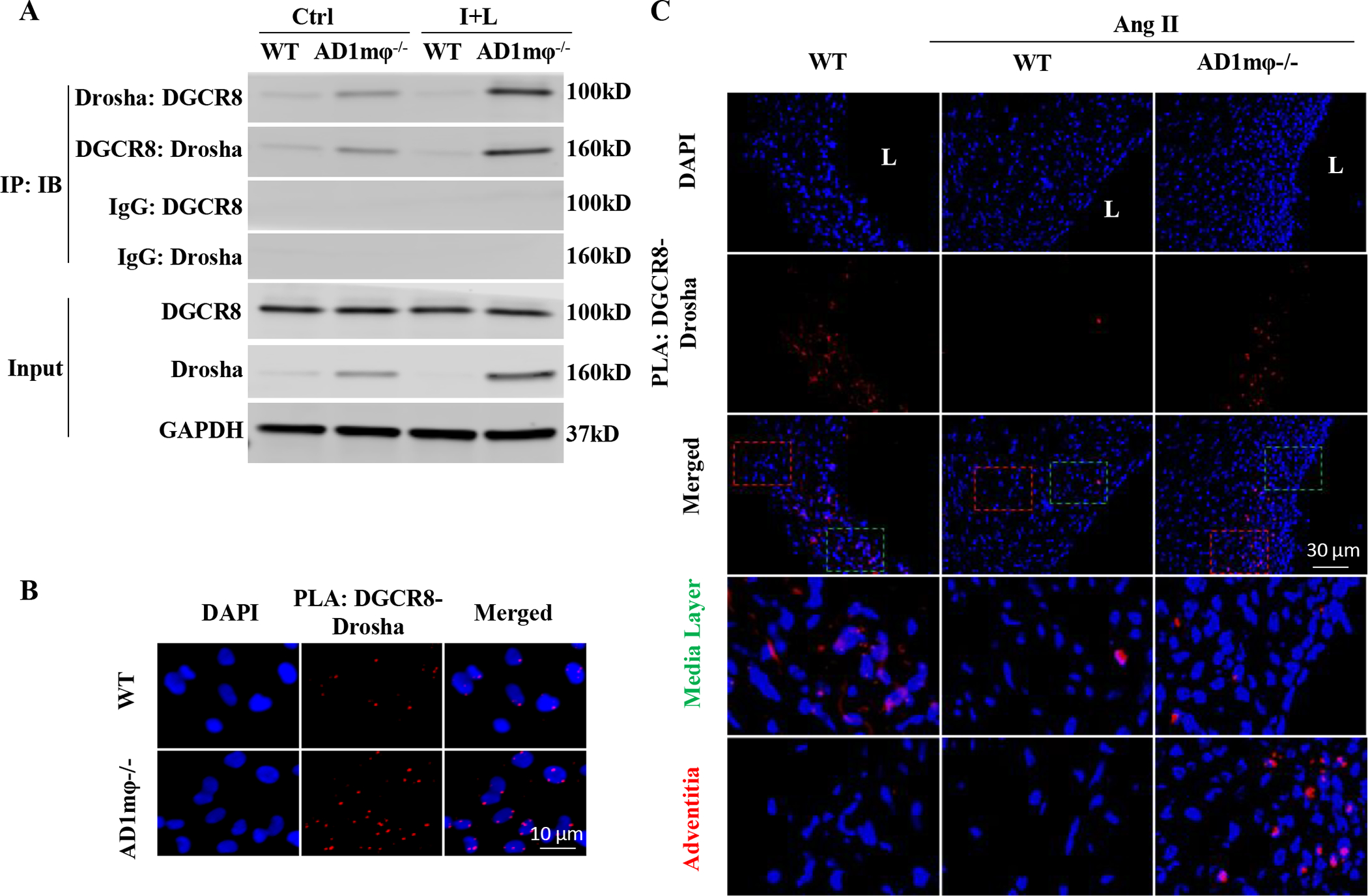
A-B, ADAR1mφ−/− (AD1mφ−/−) significantly enhanced Drosha-DGCR8 interaction in classically activated macrophages. BMDMs isolated from WT or AD6.31mφ−/− mice were treated with vehicle (Ctrl) or interferon γ (IFNγ, 100 ng/mL) and lipopolysaccharides (LPS,100 ng/mL) (I+L) for 6 h to induce macrophage classical activation. A, Coimmunoprecipitation assays were performed to detect the Drosha-DGCR8 interaction. Control (IgG), Drosha or DGCR8 antibodies were used for immunoprecipitation (IP), and immunoblotting (IB) was performed with DGCR8 and Drosha antibodies, respectively. B, In situ proximity ligation assays (PLA) were performed to confirm Drosha-DGCR8 interaction in AD1mφ−/− BMDMs activated by I+L (n=6). DAPI stains the nuclei. C, AD1mφ−/− enhanced the physical interaction between DGCR8 with Drosha in mouse AAA lesion. PLA was performed on mouse abdominal aorta or AAA sections by staining with both Drosha and DGCR8 antibodies (n=6). IgG staining was used as a negative control (online Figure XIV). DAPI stains nuclei. L: lumen. The media (green box) and adventitia areas (red box) in the merged images were enlarged in the lower panels (3.8 folds). Adventitia areas where macrophages accumulate show more Drosha-DGCR8 interaction. The quantifications of PLA signals in B and C are shown in online Figure XV.