
Figure 7: Macrophage ADAR1 and its interaction with Drosha correlate with the development of human AAA.
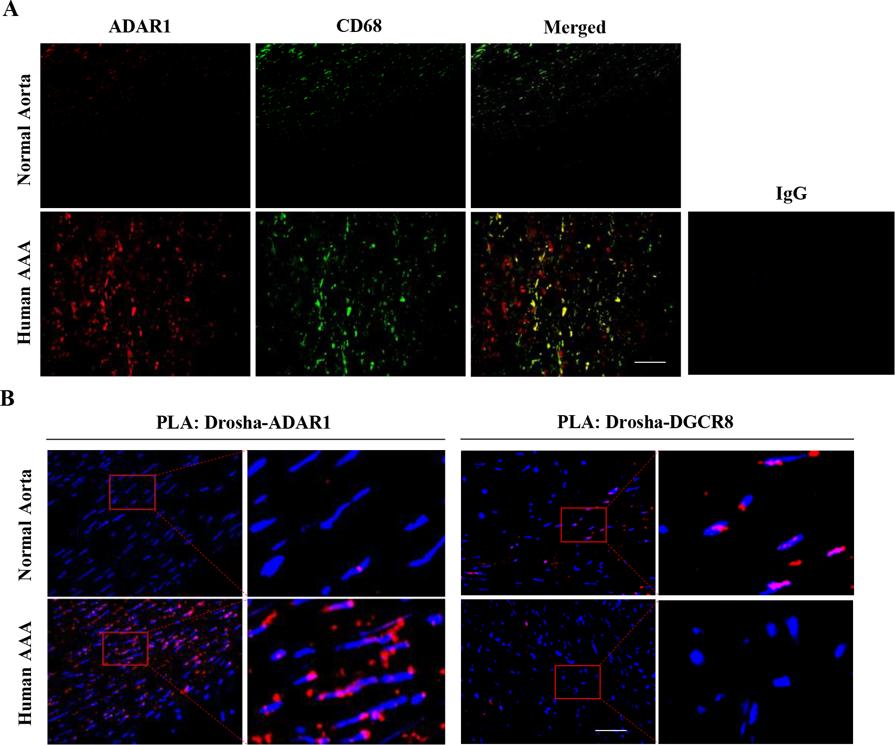
A, Normal healthy human abdominal aorta or AAA sections were co-immunostained with CD68 and ADAR1 antibodies. Red: ADAR1, Green: CD68. The areas in the red boxes were shown with a higher magnification (4.2 folds) in the left part of the panel. B, The increased ADAR1 interaction with Drosha correlated with decreased Drosha interaction with DGCR8 in human AAA lesions, as detected by In situ Duolink Proximity Ligation Assay. DAPI stains nuclei. Scale bar: 30 μm. The negative control of PLA assay is shown in online Figure XXIII. The quantitative analyses of immunostaining (A) and PLA signal (B) are shown in Online Figure XXI (n=6).