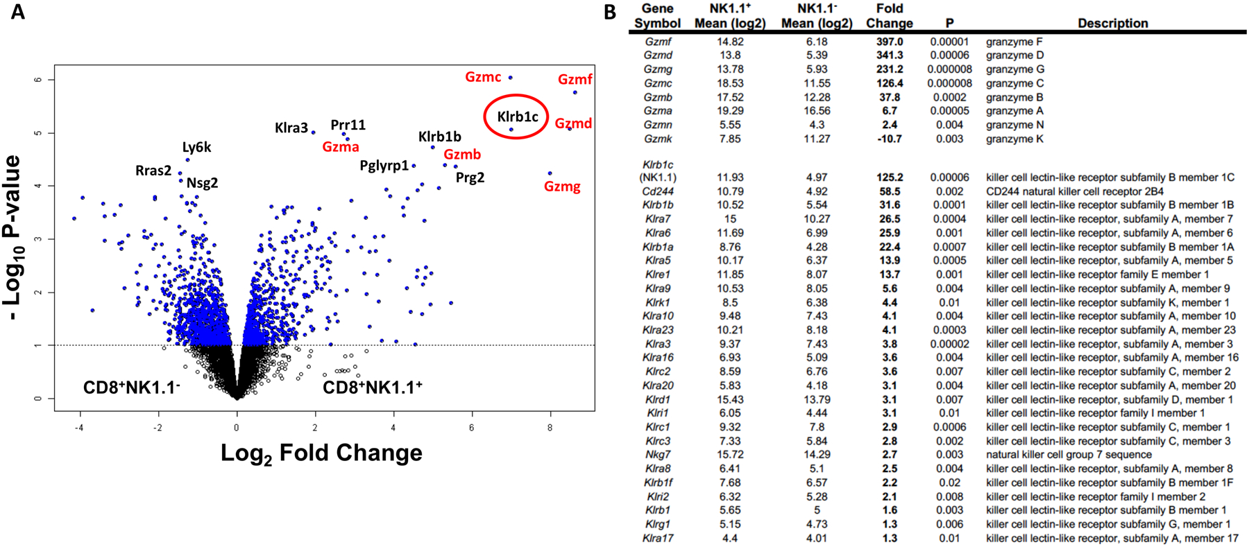
Figure 1. Gene expression analysis by microarray of antigen stimulated T cells indicates upregulated cytotoxic and innate-like characteristics among CD8+NK1.1+ cells.

A cohort of 15 mice received dendritic cell-based vaccination in combination with chemotherapy against murine pancreatic ductal adenocarcinoma. 60 days post tumor inoculation, spleens were harvested, pooled into three groups of five each and activated overnight with dendritic cells loaded with tumor antigens. Stimulated cells were then sorted by flow cytometry into NK1.1− and NK1.1+ subsets by gating on CD8+CD69+ population. (A) Volcano plot shows 1642 genes that are differentially regulated between CD8+NK1.1− and CD8+NK1.1+ cells at a univariate degree of 0.1. Top 15 genes that are differentially regulated at an FDR of 0.05 are labeled on the plot with genes from the granzyme and killer-like lectin pathways are labeled in red. NK1.1 (Klrb1c) is circled in red. (B) Fold change and P values for the genes grouped into the granzyme pathway and killer cell like receptor subfamily pathway are indicated.
